Review
. 2012 Dec 10;164(2):192-204.
doi: 10.1016/j.jconrel.2012.04.045. Epub 2012 May 18.

Opportunities and challenges for use of tumor spheroids as models to test drug delivery and efficacy

Affiliations
Review

Opportunities and challenges for use of tumor spheroids as models to test drug delivery and efficacy

Geeta Mehta et al. J Control Release. .

Abstract

Multicellular spheroids are three dimensional in vitro microscale tissue analogs. The current article examines the suitability of spheroids as an in vitro platform for testing drug delivery systems. Spheroids model critical physiologic parameters present in vivo, including complex multicellular architecture, barriers to mass transport, and extracellular matrix deposition. Relative to two-dimensional cultures, spheroids also provide better target cells for drug testing and are appropriate in vitro models for studies of drug penetration. Key challenges associated with creation of uniformly sized spheroids, spheroids with small number of cells and co-culture spheroids are emphasized in the article. Moreover, the assay techniques required for the characterization of drug delivery and efficacy in spheroids and the challenges associated with such studies are discussed. Examples for the use of spheroids in drug delivery and testing are also emphasized. By addressing these challenges with possible solutions, multicellular spheroids are becoming an increasingly useful in vitro tool for drug screening and delivery to pathological tissues and organs.


Figures

Figure 1
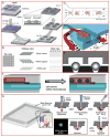
Figure 1. Spheroids are appropriate models for testing drug delivery systems in vitro.
A) In vivo, concentration of drug, oxygen and nutrients decreases on increasing distance from blood vessels. The effectiveness of drug and carrier system in vivo is a function of drug dose, potency, kinetics, molecular weight, charge, solubility in water and lipids, diffusion, barriers in the microenvironment, binding, metabolism, and sequestration. B) Spheroids are 3-D microscale tissues which exhibit an inherent gradient of nutrients, oxygen and metabolites within themselves, which leads to a central necrotic core region surrounded by quiescent viable cells and an outer layer of actively proliferating cells. Due to similarities between in vivo tissues and spheroids as well as mass transport limitations, spheroids can serve as high throughput screening platforms for drug and carrier effectiveness. Scale bar is 200 μm.
Figure 2
Figure 2. Current state-of-the-art methods of generating uniformly sized spheroids
A) A spheroid co-culture system utilizing chitosan hydrogel microarray molded with PDMS. The uniformity of spheroids generated was controlled by the patterned chitosan. Reproduced from [52] by the kind permission of Elsevier Press. B) Schematic of arrays of v-bottom PDMS microwells to generate uniformly-sized spheroids in a high-throughput fashion. Reproduced from [45] under the Creative Commons Attribution License. C) A microfluidic device with micro-posts to trap single cell suspensions into uniform clusters of cells that will then self-aggregate into spheroids. Reproduced from [49] by the kind permission of Elsevier Press. D) A microfluidic device with micro-chambers to trap single cell suspensions and induce spheroid formation. The size of the spheroids is controlled by the chamber dimensions. Reproduced from [51] by the kind permission of Springer Science. E) Cartoon of a v-bottom microwell-based device to generate spheroids of the same size. Reproduced from [44] by the kind permission of Elsevier Press. F) Schematic showing a two-layer PDMS-based microfluidic device for the generation of uniformly-sized embryoid bodies. After introducing cells into the microchannel (with surfaces blocked to prevent cell adhesion) as a uniform monolayer, cells spontaneously aggregate and break up into uniformly-sized embryoid bodies. Reproduced from [50] by permission of The Royal Society of Chemistry. (G) 384 hanging drop array plate and a cartoon of the spheroid formation process. The size of the spheroid is controlled by the number of cells seeded into each hanging drop. Reproduced from [59] by permission of The Royal Society of Chemistry.
Figure 3
Figure 3. Tissue and tumor histoids are generated through the spontaneous interaction of multiple cell types
A) Histological section of breast cancer histoid (BCH) containing BT20 cancer cells and, generated in low shear, rotating suspension culture and immunostained to demonstrate cytokeratin in cancer cells. Photographed using 20x objective; inset shows size variability. Suspension of intact, viable pancreatic tumor histoids containing fibroblasts and pancreatic cancer cells (MIAPaCa) were generated in rotating suspension cultures, and imaged under B) 10x and C) 20x objectives. Pancreatic cancer cells were transfected to express green fluorescent ZsGreen 1, a variant of the widely used GFP. These histoids can be sorted according to size and fluorescent intensity (using COPAS flow cytometer) to obtain a relatively uniform population.
Figure 4
Figure 4. Bioluminescence quantification of doxorubicin cytotoxicity in MDA-MB-231 human breast cancer cells
A) Image of luciferase activity in intact spheroids, and B) quantification of bioluminescence after 48 hours of treatment. Luminescence data were normalized to pre-treatment values for each spheroid. Loss of bioluminescence directly measures relative cytotoxicity. Error bars denote mean values + SEM.

References

    1. Han C, Wang B. Factors that impact developability of drug candidates: an overview. In: Wang B, Siahaan T, Soltero R, editors. Drug Delivery: Priciples and Applications. John Wiley & Sons; Hoboken, New Jersey: 2005. pp. 1–14.
    1. Saltzman WM. Drug Delivery: Engineering Principles for Drug Therapy. Oxford University Press; New York: 2001.
    1. Torchilin VP. Passive and active drug targeting: drug delivery to tumors as an example. In: Schafer-Korting M, editor. Drug Delivery. Springer; 2010. pp. 3–54. - PubMed
    1. Jain KK. Drug Delivery Systems- An Overview. In: Jain KK, editor. Drug Delivery Systems. Humana Press; 2008. pp. 1–50. - PubMed
    1. Karunaratne DN, Silverstein PS, Vasadani V, Young AM, Rytting E, Yops B, Audus KL. Cell culture models for drug transport studies. In: Wang B, Siahaan T, Soltero R, editors. Drug Delivery: Priciples and Applications. John Wiley & Sons; Hoboken, New Jersey: 2005. pp. 103–124.

Publication types

Substances