Clinical Trial
. 2012 Aug;36(8):1234-46.
doi: 10.1097/PAS.0b013e31825793ff.

Liver and skin histopathology in adults with acid sphingomyelinase deficiency (Niemann-Pick disease type B)

Affiliations
Clinical Trial

Liver and skin histopathology in adults with acid sphingomyelinase deficiency (Niemann-Pick disease type B)

Beth L Thurberg et al. Am J Surg Pathol. 2012 Aug.

Abstract

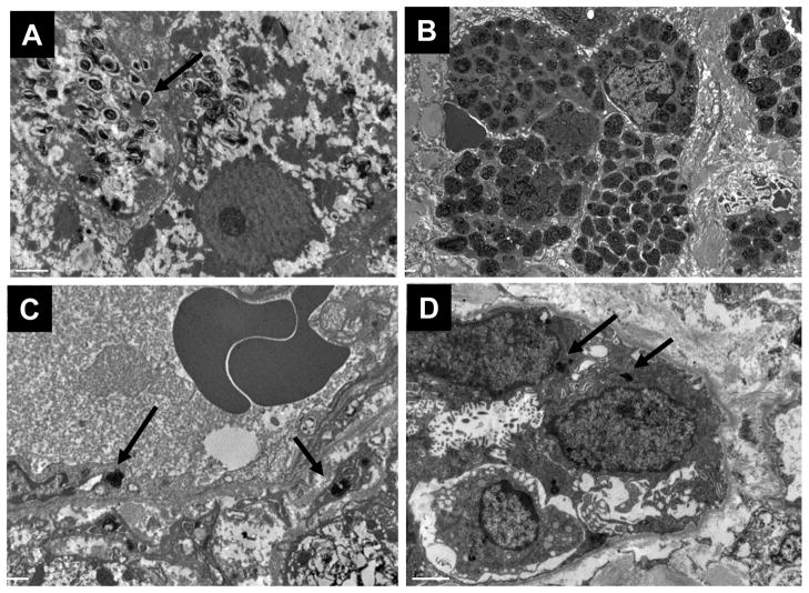
Acid sphingomyelinase deficiency (ASMD) is a lysosomal storage disorder characterized by the pathologic accumulation of sphingomyelin (SM) in multiple cell types, and occurs most prominently within the liver, spleen, and lungs, leading to significant clinical disease. Seventeen ASMD patients underwent a liver biopsy during baseline screening for a phase 1 trial of recombinant human acid sphingomyelinase (rhASM) in adults with Niemann-Pick disease type B. Eleven of the 17 were enrolled in the trial and each received a single dose of rhASM and underwent a repeat liver biopsy on day 14. Biopsies were evaluated for fibrosis, SM accumulation, and macrophage infiltration by light and electron microscopy. When present, fibrosis was periportal and pericellular, predominantly surrounding affected Kupffer cells. Two baseline biopsies exhibited frank cirrhosis. SM was localized to isolated Kupffer cells in mildly affected biopsies and was present in both Kupffer cells and hepatocytes in more severely affected cases. Morphometric quantification of SM storage in liver biopsies ranged from 4% to 44% of the microscopic field. Skin biopsies were also performed at baseline and day 14 to compare the SM distribution in a peripheral tissue with that of liver. SM storage was present at lower levels in multiple cell types of the skin, including dermal fibroblasts, macrophages, vascular endothelial cells, vascular smooth muscle cells, and Schwann cells. This phase 1 trial of rhASM in adults with ASMD provided a unique opportunity for a prospective assessment of hepatic and skin pathology in this rare disease and their potential usage as pharmacodynamic biomarkers.


Conflict of interest statement

Conflicts of Interest and Source of Funding: BLT, FOB, SR and GFC are employees of Genzyme Corporation. This study was supported by Genzyme Corporation (Cambridge, MA). MMM is the recipient of Mid-Career Patient-Oriented Research Career Development Award K24 RR021991-01 from the National Institutes of Health. The patient studies were also supported by grant 5 MO1 RR00071 for the Mount Sinai General Clinical Research Center from the National Center for Research Resources, National Institutes of Health.

Figures

Figure 1
Figure 1. Assessment of liver biopsies for fibrosis
A. Patient 6: liver biopsy with fibrosis grade of zero. Reticulin stain highlights the normal hepatocellular architecture (10× objective). B. Patient 16: liver biopsy with fibrosis grade of 4 (cirrhosis). Reticulin highlights the collapse of the hepatic sinusoidal architecture and the formation of nodules (10× objective). C. Trichrome stained section of biopsy from Patient 6. Note the absence of fibrosis and the presence of isolated, SM-laden Kupffer cells (arrows), features characteristic of mildly affected biopsies (40× objective). D. Trichrome stained section of biopsy from patient 16. Note the pericellular fibrosis which accompanies the formation of large swaths of Kupffer cells, a common feature in more severely affected biopsies (40× objective).
Figure 2
Figure 2. Sphingomyelin load and cell distribution vary amongst patient biopsies
In some biopsies, SM is present in sinusoidal Kupffer cells only (K distribution). In other biopsies, SM has accumulated in sinusoidal Kupffer cells, foci of proliferating portal Kupffer cells and hepatocytes (KH distribution). A. Patient 6: SM is present within enlarged, foamy Kupffer cells (arrows; H&E, 60× objective). B. Patient 1: SM is present within clusters of enlarged foamy Kupffer cells (K) and foamy hepatocytes (H) (H&E, 60× objective). C. In HRLM sections, SM stains purple in sinusoidal Kupffer cells (modified toluidine blue, 100× oil objective). D. In KH distribution, SM is present in both Kupffer cells and hepatocytes (modified toluidine blue, 100× oil objective). E and F. Lysenin affinity staining also highlights the differences in SM accumulation in K versus KH distribution patterns (lysenin affinity stain, 100× oil objective). G. CD68 immunohistochemistry highlights the enlarged Kupffer cells as single cells (10× objective). H. CD68 immunohistochemistry highlights enlarged Kupffer cells as single cells and as clusters proliferating in portal tracts and infiltrating bands of fibrosis (10× objective).
Figure 3
Figure 3. Electron microscopy of lysosomal sphingomyelin in liver biopsies
A. Sphingomyelin is present in hepatocytes as scattered zebra bodies and loose myelin figures (15000×). B. In Kupffer cells, SM packs the cell cytoplasm as dense myelin figures (5000×). C. Sinusoidal endothelial cells contain dense foci of SM (12000×). D. Bile duct epithelium also contains SM (8000×)
Figure 4
Figure 4. Quantification of hepatic sphingomyelin in HRLM sections by Metamorph® Analysis
A. Sphingomyelin can be quantified in liver biopsies with HRLM processing and MetaMorph® analysis. B. Metamorph® bar graph of Baseline levels of SM, solid bars. Post-infusion levels are indicated as hatched bars, when available.
Figure 5
Figure 5. Isolated liver observations
A. This image shows the typical appearance of the portal and pericellular fibrosis of the foamy cell infiltration (arrows) that is present in the majority of biopsies in this study (H&E, 40× objective). B. Trichrome stained section of same area in A highlights the fine, loose appearance of pericellular fibrosis around foamy Kupffer cells (40× objective). C. In contrast, patient 13 baseline biopsy contained dense periportal fibrosis with infiltration of lymphocytes (H&E, 40× objective). D. Trichrome stain of the same region in A shows blue staining of the dense fibrosis which is devoid of foamy cell infiltration (trichrome, 40× objective). E. Patient 10 post-treatment biopsy contains small lymphocytic foci with hepatocellular degeneration (H&E, 40× objective). F. Trichrome stain of the same region in C showing hyperchromasia in the region of hepatocellular degeneration (trichrome, 40× objective).
Figure 6
Figure 6. Sphingomyelin accumulates in multiple cell types of the skin
A. Sphingomyelin is present in fibroblasts (arrowhead) and macrophages (black arrow) of the superficial dermis (red lysenin affinity stain for SM, toluidine blue counterstain, 60× objective). B. Sphingomyelin can also be identified in endothelial cells and pericytes of capillaries in the deep dermis between adipocytes (red lysenin affinity stain for SM, toluidine blue counterstain, 60× objective). C. Electron microscopy of SM in dermal fibroblast (white arrow; 15000×). D. Electron microscopy of SM in dermal capillary endothelial cells (arrowhead) and pericytes (white arrow) (12000×). E. Electron microscopy of SM in dermal macrophage (white arrow, 25000×). F. Sphingomyelin was also present within small nerves (12000×).

References

    1. Crocker AC, Farber S. Niemann-Pick disease: a review of eighteen patients. Medicine. 1958;37(1):1–95. - PubMed
    1. Horinouchi K, Erlich S, Perl DP, et al. Acid sphingomyelinase deficient mice: a model of types A and B Niemann-Pick disease. Nature Genetics. 1995;10:288–293. - PubMed
    1. Labrune P, Bedossa P, Huguet P, et al. Fatal liver failure in two children with Niemann-Pick disease type B. J Pediatr Gastroenterol Nutr. 1991;13:104–109. - PubMed
    1. Lynch CM, Johnson J, Vaccaro C, et al. High Resolution Light Microscopy (HRLM) and Digital Analysis of Pompe Disease Pathology. J Histochem Cytochem. 2005;53 (1):63–73. - PubMed
    1. McGovern MM, Pohl-Worgall T, Deckelbaum RJ, et al. Lipid abnormalities in children with types A and B Niemann-Pick disease. J Pediatr. 2004;145:77–81. - PubMed

Publication types

Substances

LinkOut - more resources