. 2012 Aug;80(8):2905-13.
doi: 10.1128/IAI.00356-12. Epub 2012 May 21.

Neutrophils are essential for containment of Vibrio cholerae to the intestine during the proinflammatory phase of infection

Affiliations

Neutrophils are essential for containment of Vibrio cholerae to the intestine during the proinflammatory phase of infection

Jessica Queen et al. Infect Immun. 2012 Aug.

Abstract

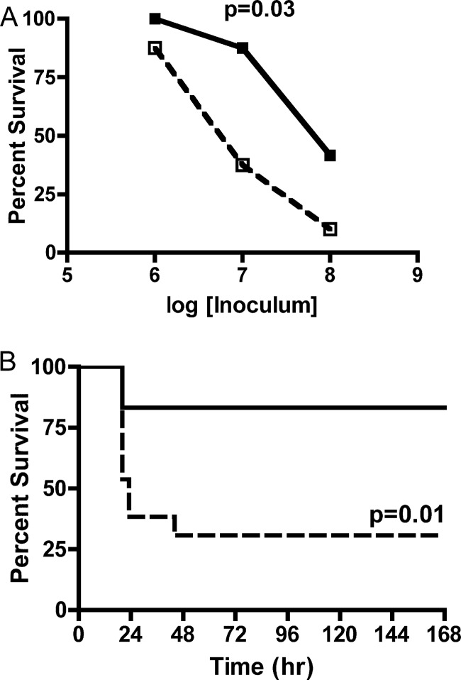
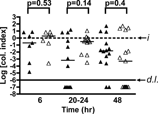
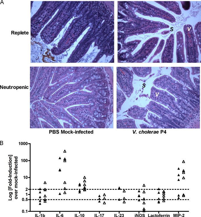
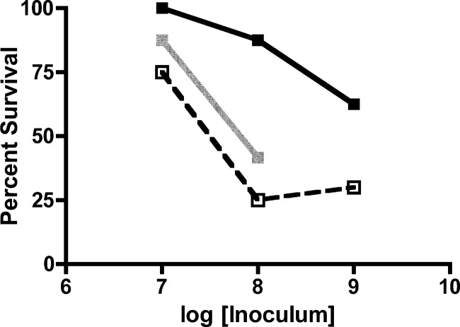
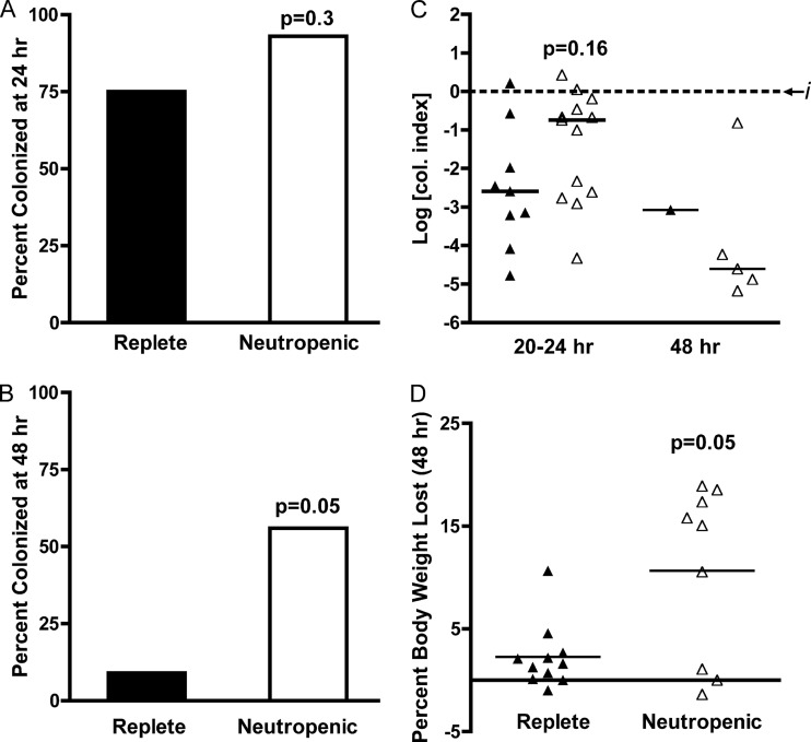
Cholera is classically considered a noninflammatory diarrheal disease, in comparison to invasive enteric organisms, although there is a low-level proinflammatory response during early infection with Vibrio cholerae and a strong proinflammatory reaction to live attenuated vaccine strains. Using an adult mouse intestinal infection model, this study examines the contribution of neutrophils to host defense to infection. Nontoxigenic El Tor O1 V. cholerae infection is characterized by the upregulation of interleukin-6 (IL-6), IL-10, and macrophage inflammatory protein 2 alpha in the intestine, indicating an acute innate immune response. Depletion of neutrophils from mice with anti-Ly6G IA8 monoclonal antibody led to decreased survival of mice. The role of neutrophils in protection of the host is to limit the infection to the intestine and control bacterial spread to extraintestinal organs. In the absence of neutrophils, the infection spread to the spleen and led to increased systemic levels of IL-1β and tumor necrosis factor alpha, suggesting the decreased survival in neutropenic mice is due to systemic shock. Neutrophils were found not to contribute to either clearance of colonizing bacteria or to alter the local immune response. However, when genes for secreted accessory toxins were deleted, the colonizing bacteria were cleared from the intestine, and this clearance is dependent upon neutrophils. Thus, the requirement for accessory toxins in virulence is negated in neutropenic mice, which is consistent with a role of accessory toxins in the evasion of innate immune cells in the intestine. Overall, these data support that neutrophils impact disease progression and suggest that neutrophil effectiveness can be manipulated through the deletion of accessory toxins.


Figures

Fig 1
Fig 1
Survival after infection with nontoxigenic V. cholerae. Mock-PBS-treated or neutrophil-depleted C57BL/6 mice were inoculated with a range of doses of nontoxigenic V. cholerae El Tor O1 strain P4, and survival was monitored over the course of 7 days. (A) Total survival of replete (solid line) or neutropenic (dotted line) mice inoculated with (1.4 to 2.1) × 106 CFU, (1.3 to 2.0) × 107 CFU, or (2.4 to 3.7) × 108 CFU. (B) Survival curve of replete (solid line) and neutropenic (dotted line) mice inoculated with (1.3 to 2.0) × 107 CFU plotted over time. The data are pooled from two experiments and analyzed by using the Fisher exact test (A) or log-rank test (B).
Fig 2
Fig 2
Small intestine colonization. Mock-PBS-treated (▲) or neutrophil-depleted (△) C57BL/6 mice were inoculated with a sublethal dose of (0.57 to 2.8) × 106 CFU of nontoxigenic V. cholerae El Tor O1 strain P4. At 6, 20 to 24, or 48 h p.i., the small intestines were collected, homogenized in PBS, and plated for CFU counting. The data for individual mice are pooled from two to three experiments per endpoint and normalized by dividing the recovered CFU by the input CFU; thus, data are plotted as the log colonization index (median shown by black line) with the dashed line at 0 representing recovered CFU that is identical to the input (i). Values below the solid line (d.l.) indicate mice colonized below the detection limit of 100 CFU in the small intestine. Indicated P values were obtained by using a two-tailed Mann-Whitney nonparametric t test comparing medians.
Fig 3
Fig 3
Intestinal histology and inflammatory gene expression. (A) Mock-PBS-treated or neutrophil-depleted C57BL/6 mice were inoculated with 1.5 × 10⁸ CFU of nontoxigenic V. cholerae El Tor O1 strain P4 or mock inoculated with PBS. At 6 h p.i., the distal 1 cm of the small intestine at the ileocecal junction was collected, fixed, embedded in paraffin, and stained with H&E. Representative ×40 images are shown, with epithelial villi (V) and sloughed cells (S) noted as marked. (B) Mock-IgG-treated or neutrophil-depleted C57BL/6 mice were inoculated with (1.5 to 2.5) × 108 CFU of nontoxigenic V. cholerae El Tor O1 strain P4 or mock-inoculated with PBS. At 6 h p.i., the distal 1 cm of the small intestine at the ileocecal junction was collected, and RNA was extracted, converted to cDNA, and used for qRT-PCR analysis of the indicated genes, compared to PBS-mock-inoculated controls, and normalized to housekeeping gene GAPDH. Dotted lines indicate 2-fold up-or-down regulation compared to PBS control mice.
Fig 4
Fig 4
Dissemination and systemic inflammation. Mock-IgG-treated or neutrophil-depleted C57BL/6 mice were inoculated with (1.4 to 4.6) × 108 CFU of nontoxigenic V. cholerae El Tor O1 strain P4 or mock-inoculated with PBS. At 6 h p.i., the small intestines (A), livers (B), spleens (C), and serum (D and E) were collected. Organs were homogenized and plated for CFU counting. Serum was analyzed for protein concentration by ELISA. The data are pooled from four experiments and analyzed by using a two-tailed Mann-Whitney nonparametric t test comparing medians (A to C) or a two-tailed Student t test (D and E).
Fig 5
Fig 5
Survival after infection with multi-toxin-deficient V. cholerae. Mock-PBS-treated or neutrophil-depleted C57BL/6 mice were inoculated with a range of doses of nontoxigenic strain KFV101 (ΔhlyA ΔrtxA ΔhapA), and survival was monitored over the course of 7 days. Total survival of replete (solid black line) or neutropenic (dotted black line) mice inoculated with (2.6 to 3.8) × 107 CFU, (3.1 to 8.4) × 108 CFU, or (3.4 to 5.2) × 109 CFU. Survival is compared to survival of replete mice inoculated with 107 and 108 doses of parent strain P4 (solid gray line), as reported in Fig. 1. The data are pooled from two experiments and analyzed by using the Fisher exact test.
Fig 6
Fig 6
Prolonged colonization after infection with multi-toxin-deficient V. cholerae. Mock-PBS-treated (filled column or triangles) or neutrophil-depleted (open column or triangles) C57BL/6 mice were inoculated with a dose of (0.6 to 2.7) × 107 CFU of nontoxigenic P4 derivative strain KFV101. At 20 to 24 h or 48 h p.i., small intestines were collected, homogenized in PBS, and plated for CFU counting. The data are pooled from two experiments per endpoint. Mice are reported as either colonized or cleared at 20 to 24 h (A) or 48 h (B), and the data were analyzed by using the Fisher exact test. The CFU in the small intestines of mice remaining colonized (C) are represented as the log colonization index (CFUrecovered/CFUinput) with the median shown (black line) and the dashed line at 0 representing recovered CFU that is identical to the input (i). The percent weight loss of mice at 48 h is indicated (D), with the mean shown (black bar). Indicated P values were obtained by using a two-tailed Mann-Whitney nonparametric t test comparing medians (C) or a two-tailed Student t test (D). Statistical analysis of median colonization index could not be completed for the 48-h time point, since only one neutrophil-replete mouse remained colonized.

References

    1. Ali M.2012. The global burden of cholera. Bull. World Health Organ. 90:209–218A - PMC - PubMed
    1. Bishop AL, Camilli A. 2011. Vibrio cholerae: lessons for mucosal vaccine design. Expert Rev. Vaccines 10:79–94 - PMC - PubMed
    1. Bromander A, Holmgren J, Lycke N. 1991. Cholera toxin stimulates IL-1 production and enhances antigen presentation by macrophages in vitro. J. Immunol. 146:2908–2914 - PubMed
    1. Bromander AK, Kjerrulf M, Holmgren J, Lycke N. 1993. Cholera toxin enhances alloantigen presentation by cultured intestinal epithelial cells. Scand. J. Immunol. 37:452–458 - PubMed
    1. Bubshait SA, Al-Turki K, Qadri MH, Fontaine RE, Cameron D. 2000. Seasonal, nontoxigenic Vibrio cholerae O1 Ogawa infections in the eastern region of Saudi Arabia. Int. J. Infect. Dis. 4:198–202 - PubMed

Publication types