. 2012 Jun 26;109(26):10218-23.
doi: 10.1073/pnas.1118448109. Epub 2012 May 21.

Direct chemical evidence for eumelanin pigment from the Jurassic period

Affiliations

Direct chemical evidence for eumelanin pigment from the Jurassic period

Keely Glass et al. Proc Natl Acad Sci U S A. .

Abstract

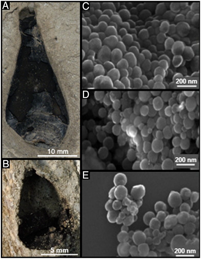
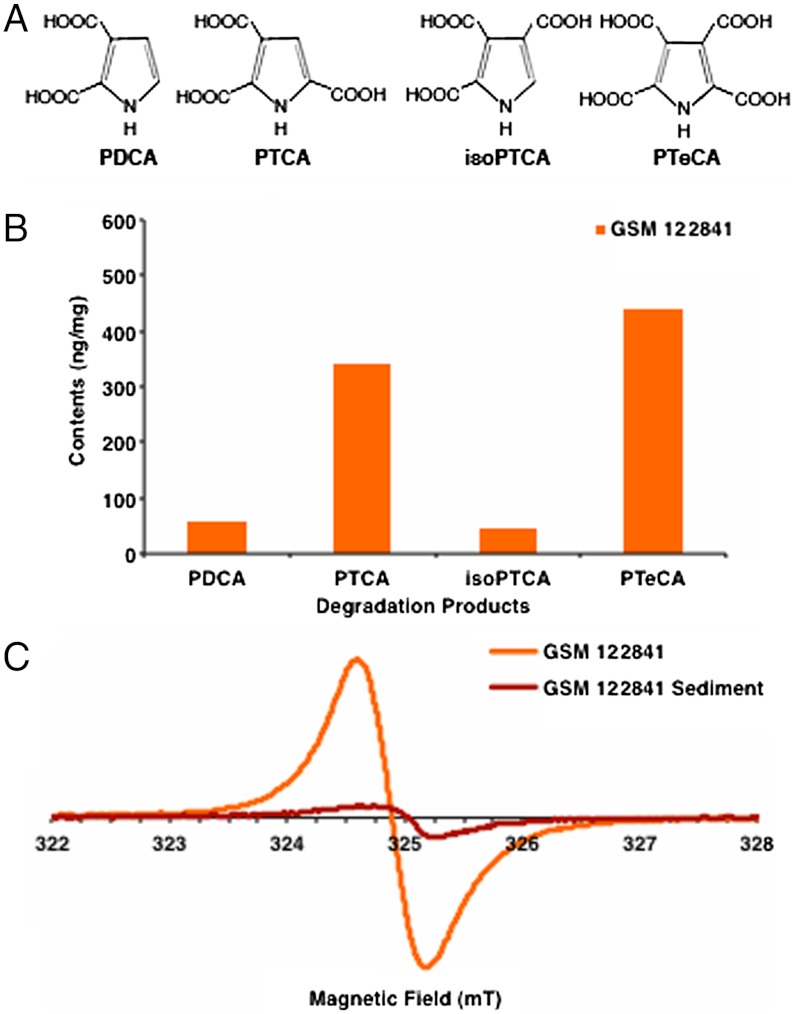
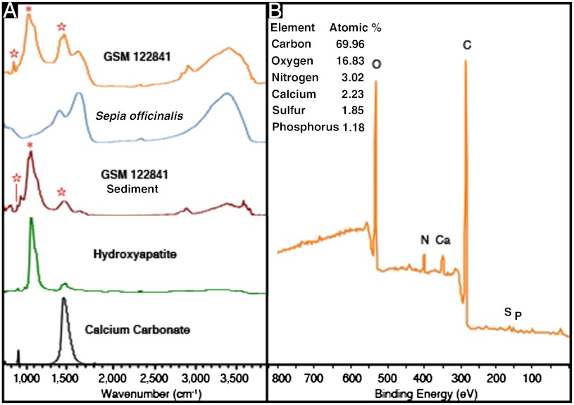
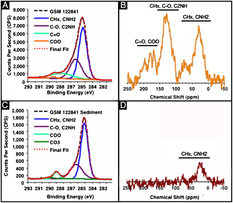
Melanin is a ubiquitous biological pigment found in bacteria, fungi, plants, and animals. It has a diverse range of ecological and biochemical functions, including display, evasion, photoprotection, detoxification, and metal scavenging. To date, evidence of melanin in fossil organisms has relied entirely on indirect morphological and chemical analyses. Here, we apply direct chemical techniques to categorically demonstrate the preservation of eumelanin in two > 160 Ma Jurassic cephalopod ink sacs and to confirm its chemical similarity to the ink of the modern cephalopod, Sepia officinalis. Identification and characterization of degradation-resistant melanin may provide insights into its diverse roles in ancient organisms.


Conflict of interest statement

The authors declare no conflict of interest.

Figures

Fig. 1.
Fig. 1.
Photographs of intact Jurassic coleoid ink sac specimens GSM 122841 (A) and GSM 120386 (B). SEM images of S. officinalis melanin (C), GSM 122841 (D), and GSM 120386 (E). All SEM images were taken at the same magnification. The average size of the spherical structures shown in CE is 140 ± 20 nm, 168 ± 30 nm, and 156 ± 30 nm, respectively, where ± indicates the standard deviation.
Fig. 2.
Fig. 2.
(A) The molecular structure of the unique melanin markers. (B) Quantification of melanin markers produced by chemical degradation of fossil specimen GSM 122841. Degradation of the sediment of GSM 122841 yielded 1.7 ng/mg PDCA, 2.2 ng/mg PTCA, < 2.2 ng/mg isoPTCA, and < 1.3 ng/mg of PTeCA (Table S1). (C) The EPR spectra of GSM 122841 and its sediment exhibited the linewidth and g-factor characteristic of eumelanin. The same quantity of sample pigment and sediment were used to provide a qualitative comparison of the amount of eumelanin present.
Fig. 3.
Fig. 3.
(A) IR absorption spectra for GSM 122841, S. officinalis melanin, GSM 122841 sediment, and standards of hydroxyapatite and calcium carbonate. The absorption bands marked with asterisk and stars are attributed to phosphate and carbonate group, respectively. (B) XPS scan of GSM 122841, revealing the major elements present in the top 5–10 nm of the fossil specimen. Additional elements present in greater than 0.1 atomic % (the detection limit of the instrument) include: silicon 2.70, aluminum 2.04, and fluorine 0.18.
Fig. 4.
Fig. 4.
(A) High-resolution carbon XPS scan of GSM 122841, (B) CP-MAS SSNMR spectrum of GSM 122841, (C) high-resolution carbon XPS scan of GSM 122841 sediment, and (D) CP-MAS SSNMR spectrum of GSM 122841 sediment. (A) The average percent and standard deviation of the functional groups present in the top 5–10 nm of GSM 122841 sample are, as follows: 54 ± 1, 32 ± 3, 13 ± 4, and 1 ± 1% for CHx, CNH2; C─O, C2NH; C═O; and COO based on fitting with mixed Gaussian (30%) Lorentzian peaks. (B) The presence of the functional groups revealed in the XPS data is confirmed for the bulk sample through its SSNMR spectrum. (C) The average percent and standard deviation of the functional groups present in the top 5–10 nm of GSM 122841 sediment are: 65 ± 3, 26 ± 2, 6 ± 1, and 3 ± 1% for CHx, CNH2; C─O, C2NH; COO; and CO3 based on fitting with mixed Gaussian (30%) Lorentzian peaks. No C═O peak was present in the GSM 122841 sediment. (D) The result of the XPS analysis of the GSM 122841 sediment is corroborated by the lack of key melanin aromatic and carbonyl signals in its SSNMR spectrum.

References

    1. Schweitzer MH. Soft tissue preservation in terrestrial mesozoic vertebrates. Annu Rev Earth Planet Sci. 2011;39:187–216.
    1. Briggs DEG, Evershed RP, Lockheart MJ. The biomolecular paleontology of continental fossils. Paleobiology. 2000;26:169–193.
    1. Ito S. High-performance liquid-chromatography (HPLC) analysis of eu- and pheomelanin in melanogenesis control. J Invest Dermatol. 1993;100:S166–S171. - PubMed
    1. Simon JD, Peles DN. The red and the black. Acc Chem Res. 2010;43:1452–1460. - PubMed
    1. Dadachova E, et al. The radioprotective properties of fungal melanin are a function of its chemical composition, stable radical presence and spatial arrangement. Pigment Cell Melanoma Res. 2008;21:192–199. - PubMed

Publication types

MeSH terms

LinkOut - more resources