. 2012 Jun 5;109(23):9149-54.
doi: 10.1073/pnas.1119449109. Epub 2012 May 21.

Full-length axon regeneration in the adult mouse optic nerve and partial recovery of simple visual behaviors

Affiliations

Full-length axon regeneration in the adult mouse optic nerve and partial recovery of simple visual behaviors

Silmara de Lima et al. Proc Natl Acad Sci U S A. .

Erratum in

  • Proc Natl Acad Sci U S A. 2012 Aug 14;109(33):13465

Abstract

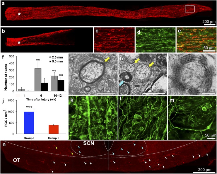
The mature optic nerve cannot regenerate when injured, leaving victims of traumatic nerve damage or diseases such as glaucoma with irreversible visual losses. Recent studies have identified ways to stimulate retinal ganglion cells to regenerate axons part-way through the optic nerve, but it remains unknown whether mature axons can reenter the brain, navigate to appropriate target areas, or restore vision. We show here that with adequate stimulation, retinal ganglion cells are able to regenerate axons the full length of the visual pathway and on into the lateral geniculate nucleus, superior colliculus, and other visual centers. Regeneration partially restores the optomotor response, depth perception, and circadian photoentrainment, demonstrating the feasibility of reconstructing central circuitry for vision after optic nerve damage in mature mammals.


Conflict of interest statement

The authors declare no conflict of interest.

Figures

Fig. 1.
Fig. 1.
Optic nerve regeneration. (A and B) Ten weeks after optic nerve injury, CTB+ axons (red) extend the full length of the optic nerve in Group I mice (A) but only part-way in Group II (B). (C–E) Region in the white rectangle of A double-labeled for CTB and GAP-43. (F) Time-course of axon regeneration in Group I mice as visualized by CTB-labeling. Bars show numbers of CTB+ axons counted 2.5 mm (gray) and 5 mm (black) from the injury site at 1, 6, and 10 wk after nerve injury. Note progressive increase in lengthy axons over time. (G–I) High-power EM images through optic nerves of Group I mice show axons that appear to be undergoing remyelination (G, arrow), unmyelinated axons (yellow arrows), and axons with thick myelin (blue arrow, H) that are seen at higher magnification to contain multiple lamellae of tightly-packed myelin (I). (J–M) RGC survival. (J) Average number of viable RGCs counted in the retinas of mice in Groups I and II. (K–M) RGC survival visualized by immunostaining for βIII-tubulin in uninjured (K), Group I (l), and Group II (M) mice. (N) CTB+ axons in the optic chiasm. Note axons coursing in the optic tract on the right side (contralateral to the regenerating optic nerve) and a smaller number on the left side (white arrowheads). Some CTB+ profiles are seen within the SCN bilaterally (blue arrowheads) and some are outside this area (yellow arrowheads). Error bars in F and J show SEM. **P < 0.01; ***P < 0.001. (Scale bars: A and B, 200 μm; CE, 50 μm; KM, 50 μm. All others as indicated.)
Fig. 2.
Fig. 2.
Reinnervation of the lateral geniculate nucleus. CTB+ fibers at the rostral (A), middle (B), and caudal (C) levels of the DLG contralateral to the regenerating optic nerve in a Group I mouse. Counterstaining for the neuronal protein NeuN shows CTB+ fibers to be confined to the neuropil of the DLG (dotted line). (E) CTB+ axons are absent in the DLG ipsilateral to the regenerating nerve in the same mouse shown in A–D. (F) No CTB+ fibers are seen in the DLG of any Group II mice. (G–I) Reinnervation of the VLG contralateral to the regenerating optic nerve. CTB+ axons (G) show intense labeling for the RGC-specific marker ERRβ (H and I), and colocalize with the presynaptic marker Vglut2 (J). (K and L) Distribution of the postsynaptic marker PSD-95 at low (K) and high magnification (L). L is a single confocal plane showing apposition of CTB+ terminals and PSD-95+ structures. Side panels show z-stacks of images in the orthogonal planes. (Scale bars: A–I, 100 μm; all others, 20 μm.)
Fig. 3.
Fig. 3.
Reinnervation of other target areas. (A–D, F, and G) Superior colliculus. CTB+ axons are seen at the rostral (A), middle (B), and caudal (C) levels of the SC of a Group I mouse. (D) Counterstaining with an antibody to NeuN shows that CTB+ axons are within the neuropil of the SC. Dotted line indicates approximate ventral extent of the SC. Absence of CTB+ axons in the SC across from the side shown in A–D (E) and in a Group II mouse (F). G–L Reinnervation of the OPT and MTN. CTB+ fibers are abundant on the side contralateral to the regenerating optic nerve (G and J) but are absent from the ipsilateral side (H and K) and from either side in Group II mice I and L. (Scale bars: 100 μm. Bar in C is for panels AC, E, and F; bar in I is for GI; bar in L is for JL).
Fig. 4.
Fig. 4.
Partial recovery of visually guided behaviors. (A) Top-down view of visual cliff. (B) Average latency to step off shallow end (Left) and total time spent on shallow end (Right). ***P < 0.01. (C) Population frequency histogram for time spent on shallow end. (D) Top-down view of apparatus used to evaluate OMR. (E) Average OMR (response threshold, cycles/degree) as a function of time. (F) Frequency distribution of the OMR. Note that y-axes in E and F are discontinuous. (G) Circadian photoentrainment: percentage of overall activity in 1-h bins for individual mice (Left) and group averages (Right). Mice were maintained on a continuous cycle of lights on at 7:00 AM and off at 7:00 PM before testing and for the first 2.5 d in the activity monitor. The light cycle was set back 6 h on day 3. Error bars represent SEM.

Comment in

References

    1. Benowitz LI, Yin Y. Optic nerve regeneration. Arch Ophthalmol. 2010;128:1059–1064. - PMC - PubMed
    1. Sun F, et al. Sustained axon regeneration induced by co-deletion of PTEN and SOCS3. Nature. 2011;480:372–375. - PMC - PubMed
    1. Yin Y, et al. Oncomodulin is a macrophage-derived signal for axon regeneration in retinal ganglion cells. Nat Neurosci. 2006;9:843–852. - PubMed
    1. Yin Y, et al. Oncomodulin links inflammation to optic nerve regeneration. Proc Natl Acad Sci USA. 2009;106:19587–19592. - PMC - PubMed
    1. Kurimoto T, et al. Long-distance axon regeneration in the mature optic nerve: Contributions of oncomodulin, cAMP, and pten gene deletion. J Neurosci. 2010;30:15654–15663. - PMC - PubMed

Publication types

MeSH terms