. 2012 Jun 5;109(23):8931-6.
doi: 10.1073/pnas.1200874109. Epub 2012 May 21.

Structural basis for homeodomain recognition by the cell-cycle regulator Geminin

Affiliations

Structural basis for homeodomain recognition by the cell-cycle regulator Geminin

Bo Zhou et al. Proc Natl Acad Sci U S A. .

Abstract

Homeodomain-containing transcription factors play a fundamental role in the regulation of numerous developmental and cellular processes. Their multiple regulatory functions are accomplished through context-dependent inputs of target DNA sequences and collaborating protein partners. Previous studies have well established the sequence-specific DNA binding to homeodomains; however, little is known about how protein partners regulate their functions through targeting homeodomains. Here we report the solution structure of the Hox homeodomain in complex with the cell-cycle regulator, Geminin, which inhibits Hox transcriptional activity and enrolls Hox in cell proliferative control. Side-chain carboxylates of glutamates and aspartates in the C terminus of Geminin generate an overall charge pattern resembling the DNA phosphate backbone. These residues provide electrostatic interactions with homeodomain, which combine with the van der Waals contacts to form the stereospecific complex. We further showed that the interaction with Geminin is homeodomain subclass-selective and Hox paralog-specific, which relies on the stapling role of residues R43 and M54 in helix III and the basic amino acid cluster in the N terminus. Interestingly, we found that the C-terminal residue Ser184 of Geminin could be phosphorylated by Casein kinase II, resulting in the enhanced binding to Hox and more potent inhibitory effect on Hox transcriptional activity, indicating an additional layer of regulation. This structure provides insight into the molecular mechanism underlying homeodomain-protein recognition and may serve as a paradigm for interactions between homeodomains and DNA-competitive peptide inhibitors.


Conflict of interest statement

The authors declare no conflict of interest.

Figures

Fig. 1.
Fig. 1.
C terminus of Geminin specifically interacts with Hoxc9. (A) Hoxc9-HD was pulled down by GST-tagged Gem-FL and its deletion mutants, and visualized by Coomassie blue staining. (B) The defined region of Geminin required for Hoxc9-HD interaction. Hoxc9-HD was pulled down by GST-tagged fragments of Geminin C terminus and visualized by Coomassie blue staining. (C) Part of the 1H-15N HSQC spectra of 15N-labeled Hoxc9-HD in free form (red) overlaid with that titrated with nonlabeled Gem-HBR at a molar ratio of 1:2 (blue). (D) Part of the 1H-15N HSQC spectra of 15N-labeled Gem-HBR in free form (red) overlaid with that titrated with nonlabeled Hoxc9-HD at a molar ratio of 1:2 (blue). (E) ITC measurement of Hoxc9-HD binding to Gem-HBR. (F) Schematic illustration of reported and currently identified protein binding motifs on Geminin.
Fig. 2.
Fig. 2.
Three-dimensional structure of Hoxc9-HD/Gem-HBR complex. (A) Ribbon diagram of a representative structure from the ensemble. Hoxc9-HD is colored in magenta, and Gem-HBR is depicted in green. (B) The complex shown with Gem-HBR in ribbon and Hoxc9-HD in surface representation. (C) Expanded view of the complex binding interface. Geminin residues are labeled in red and Hox residues in black. (D) The complex is illustrated by surface representation for Hoxc9-HD and by stick model for Gem-HBR (blue: basic residues; cyan: polar residues; green: hydrophobic residues; gray: noninteracting residues).
Fig. 3.
Fig. 3.
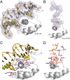
Structural comparison of Antp-HD/DNA and Hoxc9-HD/Gem-HBR complexes. Structural alignment of homeodomains in complexes of Antp-HD/DNA (A, PDB: 1AHD) and Hoxc9-HD/Gem-HBR (B). Comparison of detailed structural features of homeodomain binding interfaces between DNA (C) and Gem-HBR (D). Protein residues are labeled as described in Fig. 2C.
Fig. 4.
Fig. 4.
Effects of wild-type and mutated Geminin on Hox-DNA binding and Hox-induced transcription of the reporter gene. (A) Inhibition of Hoxc9-DNA binding. EMSA was performed using GST-tagged Hoxc9-FL (2 μg) and increasing amount (10, 25, and 60 μg) of GB1-tagged Gem-HBR or Gem-HBRmt6. The SDS/PAGE Coomassie staining gel of the input proteins is shown in Fig. S5B. (B) Inhibition of Hoxd9-mediated transcriptional activation. Gem-FL or Gem-FLmt6 alone transfected with pTHCR did not alter the reporter activity (bars 7 and 8). Gem-FL cotransfected with Hoxd9 suppressed the luciferase activity induced by Hoxd9 (bars 3 and 4), whereas no repression was observed when Gem-FLmt6 was cotransfected with Hoxd9 (bars 5 and 6). Results are presented as mean ± SEM and obtained from three independent experiments performed in triplicates. Data were analyzed by one-way ANOVA followed with Newman–Keuls multiple comparison test. **P < 0.01. (C) Comparison of Gem-FL wild-type (WT) and S184E mutant in inhibition of Hoxd9-mediated transcriptional activation. Increasing amount of Gem-FL and Gem-FL S184E were cotransfected with Hoxd9. Percent luciferase activity is the relative light units detected from the luciferase enzyme assay in cotransfected cells relative to control cells transfected with Hoxd9 alone. Results are presented as mean ± SEM and obtained from four independent experiments each performed in duplicates. Data were analyzed by Student t test. *P < 0.05. **P < 0.01 significantly different between wild-type and mutant.
Fig. 5.
Fig. 5.
Differential binding affinity of homeodomain subclasses and Hox paralogs to Geminin. (A) Sequence alignment of homeodomains of Hox paralogs and various subclasses. Residues Trp48, Phe49, Asn51, and Arg53, which are conserved across all homeodomains with major implications for DNA binding and overall stability of the homeodomain structure, are highlighted in green. Residues mainly contributing to the differential binding affinity with Geminin are highlighted in yellow. Residue numbering within the homeodomain is shown above and in the full-length protein is shown on the right. (B) A schematic illustration for the coordination of cell differentiation and proliferation through mutual regulation of posterior Hox proteins and Geminin. The C terminus of Geminin interacts with the homeodomain to inhibit Hox-induced gene transcription and cell differentiation; Hox prevents Geminin from binding to Cdt1, possibly through steric hindrance, and subsequently facilitates DNA synthesis and cell proliferation. CK2 could phosphorylate S184 at the C terminus of Geminin and enhance its binding with Hox. Furthermore, homeodomains of six subclasses (Pax6, Six3, Oct4, ISL1, Msx1, Hox) and three Hox paralogs (Hoxb1, Hoxc9, Hoxd10) have different binding affinity to Gem-HBR, in which posterior Hox proteins (Hoxc9, Hoxd10) showed the strongest binding. Residues that mainly account for the binding constant difference are the N-terminal basic amino acids at positions 2–5 (each represented by a “+”) and amino acids at positions 43 and 54 in helix III (shown with side-chain cartoon).

References

    1. Gehring WJ. Homeo boxes in the study of development. Science. 1987;236:1245–1252. - PubMed
    1. Gehring WJ, Affolter M, Bürglin T. Homeodomain proteins. Annu Rev Biochem. 1994;63:487–526. - PubMed
    1. Krumlauf R. Hox genes in vertebrate development. Cell. 1994;78:191–201. - PubMed
    1. Wellik DM. Hox genes and vertebrate axial pattern. Curr Top Dev Biol. 2009;88:257–278. - PubMed
    1. Banerjee-Basu S, Baxevanis AD. Molecular evolution of the homeodomain family of transcription factors. Nucleic Acids Res. 2001;29:3258–3269. - PMC - PubMed

Publication types

Associated data