. 2012 Jun 5;109(23):9059-64.
doi: 10.1073/pnas.1110620109. Epub 2012 May 21.

Release of dendritic cells from cognate CD4+ T-cell recognition results in impaired peripheral tolerance and fatal cytotoxic T-cell mediated autoimmunity

Affiliations

Release of dendritic cells from cognate CD4+ T-cell recognition results in impaired peripheral tolerance and fatal cytotoxic T-cell mediated autoimmunity

Sabine Muth et al. Proc Natl Acad Sci U S A. .

Abstract

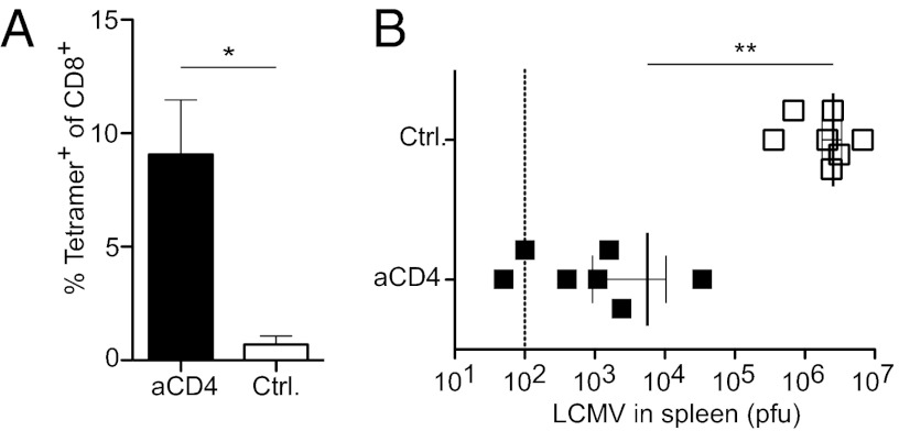
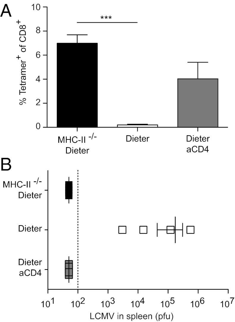
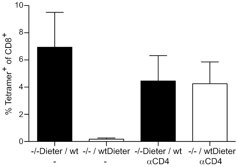
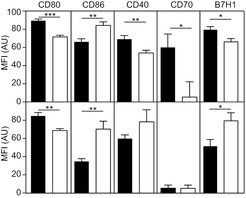
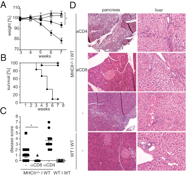
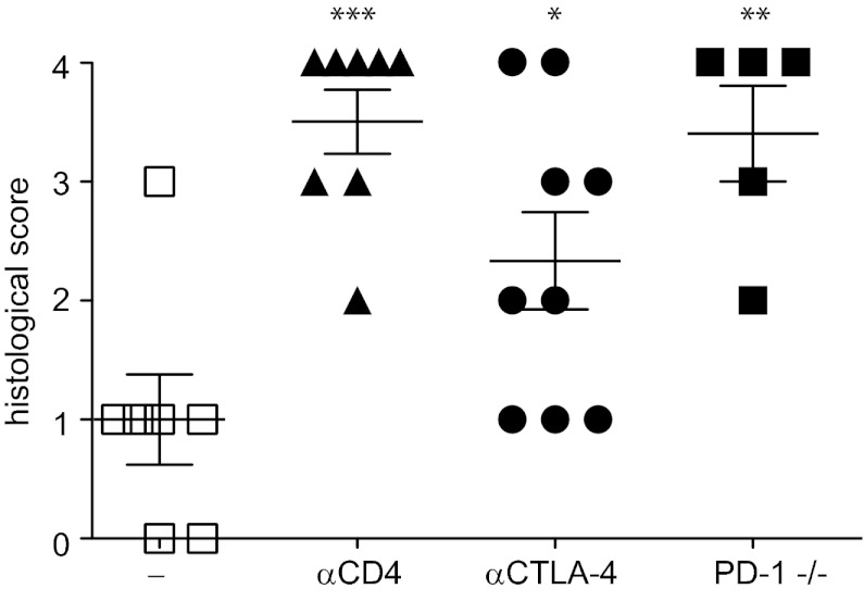
Resting dendritic cells (DCs) induce tolerance of peripheral T cells that have escaped thymic negative selection and thus contribute significantly to protection against autoimmunity. We recently showed that CD4(+)Foxp3(+) regulatory T cells (Tregs) are important for maintaining the steady-state phenotype of DCs and their tolerizing capacity in vivo. We now provide evidence that DC activation in the absence of Tregs is a direct consequence of missing DC-Treg interactions rather than being secondary to generalized autoimmunity in Treg-less mice. We show that DCs that lack MHC class II and thus cannot make cognate interactions with CD4(+) T cells are completely unable to induce peripheral CD8(+) T-cell tolerance. Consequently, mice in which interactions between DC and CD4(+) T cells are not possible develop spontaneous and fatal cytotoxic T lymphocyte-mediated autoimmunity.


Conflict of interest statement

The authors declare no conflict of interest.

Figures

Fig. 1.
Fig. 1.
Steady-state DCs induce priming of functional CTLs instead of tolerance after CD4 depletion. DIETER mice were injected i.v. with 0.5 mg of GK1.5 (closed symbols) or isotype control (open symbols) on day −1. On day 0, antigen presentation was induced by injection of 2 mg of tamoxifen i.p. (A) On day 8, expansion of GP33-41 specific CTLs was detected in the blood by staining with MHC class I tetramers. (B) On day 8, mice were challenged with 200 pfu of LCMV-WE i.v., and virus titers in the spleen were determined on day 13. n = 7 per group. Results are representative of four independent experiments.
Fig. 2.
Fig. 2.
Antigen presentation on MHC class II-deficient DCs results in functional CTL priming instead of tolerance induction. Mixed irradiation BM chimeric mice that had been reconstituted with equal numbers of MHC-II−/− DIETER + WT (CD45.1) (closed bars) or equal numbers of DIETER + WT (CD45.1) (open bars) BM cells were i.p. injected with 2 mg of tamoxifen on day 0 to induce antigen presentation on DCs. As controls, DIETER + WT mixed chimeric mice were depleted of CD4+ T cells on day −1. (A) On day 8, expansion of GP33-41–specific CTLs was detected in the blood by staining with MHC class I tetramers. (B) On day 8, mice were challenged with 200 pfu of LCMV-WE i.v., and virus titers in the spleen were measured on day 13. **P < 0.01. n = 4 per group. Results are representative of four independent experiments.
Fig. 3.
Fig. 3.
Normal numbers of FoxP3+ CD4+ regulatory T cells in mice lacking MHC class II expression on half their APCs. Frequencies of FoxP3+ CD4+ T cells among all lymphocytes in WT + WT (Upper) and MHC class II−/−/WT (Lower) mixed BM chimeric mice were determined by flow cytometry at 7 wk after reconstitution. Data are mean (SEM). n = 5.
Fig. 4.
Fig. 4.
Loss of tolerizing capacity by MHC class II-deficient, but not MHC class II-competent, DCs in mice lacking MHC class II expression on half of their APCs. Mixed irradiation BM chimeric mice that had been reconstituted with equal numbers of MHC-II−/−DIETER and WT (CD45.1) BM cells (closed bars) or equal numbers of MHC class II−/− and WT DIETER BM cells (open bars) were injected with 2 mg of tamoxifen i.p. on day 0 to induce antigen presentation on DCs. Some mice had been depleted of CD 4+ T cells on day −1. On day 8, expansion of LCMV GP33-41–specific CD8+ T cells was detected in the blood by staining with MHC class I tetramers. n = 5 per group. Results are representative of two independent experiments.
Fig. 5.
Fig. 5.
MHC II-deficient DCs are activated compared with WT DCs. Expression of CD80, CD86, CD40, CD70, and B7-H1 on CD45.1+ (WT, open bars) and CD45.1 (MHC class II−/−, filled bars) DCs isolated from spleen (Upper) or pooled peripheral lymph nodes (Lower) of MHC class II−/− + WT (CD45.1) mixed BM chimeric mice was quantified by flow cytometry. *P < 0.05; **P < 0.01; ***P < 0.001. Median fluorescence intensities of 10 mice are shown. Results are representative of three independent experiments.
Fig. 6.
Fig. 6.
Release of APCs from CD4+ T-cell control results in autoimmunity caused by CD8+ T cells. MHC-II−/− + WT mixed BM chimeric mice (closed symbols) and WT + WT control chimeric mice (open symbols) were either left untreated (squares) or depleted of CD8+ T cells (triangles) or CD4+ cells (circles) starting at 3 wk after reconstitution. (A) Body weight was measured weekly over 10 wk after BM reconstitution. (B) Survival of mixed BM chimeric mice after BM reconstitution. (C) Disease score (posture, weight loss, activity, fur texture, and skin integrity) of mixed BM chimeric mice was determined at 7 wk after BM reconstitution. (D) Histological analysis of pancreas and liver of untreated or CD4-depleted MHC-II−/− + WT and WT + WT mixed BM chimeric mice at 7 wk after reconstitution.
Fig. 7.
Fig. 7.
PD-1 and CTLA-4 are involved in suppression of CD8+ T-cell–mediated autoimmunity. MHC-II−/− + WT mixed BM chimeric mice and MHC-II−/− + PD-1−/− chimeric mice were either left untreated (squares) or depleted of CD4+ T cells (triangles) or treated with a monoclonal antibody blocking CTLA-4 (circles) starting at 3 wk after reconstitution. Comparisons with the untreated MHC-II−/− + WT group: P < 0.05; *P < 0.01; ***P < 0.001. At 6 wk after reconstitution, mononuclear infiltration to the pancreas was scored on H&E-stained sections.

References

    1. Hawiger D, et al. Dendritic cells induce peripheral T cell unresponsiveness under steady-state conditions in vivo. J Exp Med. 2001;194:769–779. - PMC - PubMed
    1. Bonifaz L, et al. Efficient targeting of protein antigen to the dendritic cell receptor DEC-205 in the steady state leads to antigen presentation on major histocompatibility complex class I products and peripheral CD8+ T cell tolerance. J Exp Med. 2002;196:1627–1638. - PMC - PubMed
    1. Probst HC, Lagnel J, Kollias G, van den Broek M. Inducible transgenic mice reveal resting dendritic cells as potent inducers of CD8+ T cell tolerance. Immunity. 2003;18:713–720. - PubMed
    1. Garbi N, Hämmerling GJ, Probst HC, van den Broek M. Tonic T cell signaling and T cell tolerance as opposite effects of self-recognition on dendritic cells. Curr Opin Immunol. 2010;22:601–608. - PubMed
    1. Probst HC, McCoy K, Okazaki T, Honjo T, van den Broek M. Resting dendritic cells induce peripheral CD8+ T cell tolerance through PD-1 and CTLA-4. Nat Immunol. 2005;6:280–286. - PubMed

Publication types

LinkOut - more resources