. 2012 Aug 15;590(16):3821-39.
doi: 10.1113/jphysiol.2011.225987. Epub 2012 May 21.

Requirement of neuronal connexin36 in pathways mediating presynaptic inhibition of primary afferents in functionally mature mouse spinal cord

Affiliations

Requirement of neuronal connexin36 in pathways mediating presynaptic inhibition of primary afferents in functionally mature mouse spinal cord

Wendy Bautista et al. J Physiol. .

Abstract

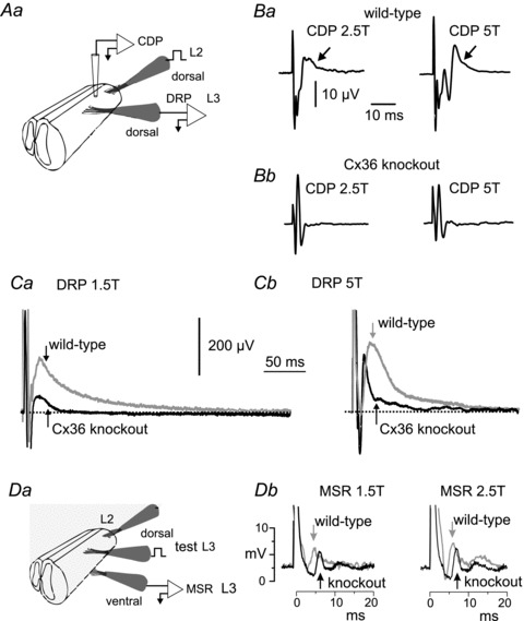
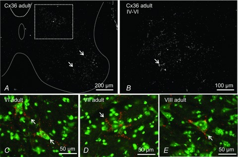
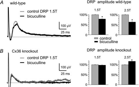
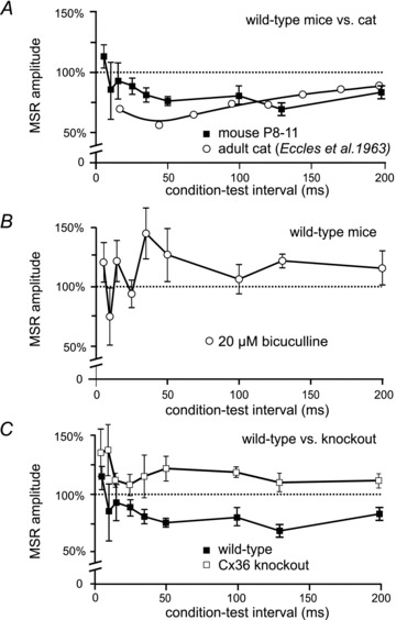
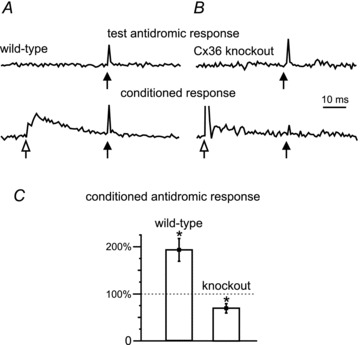
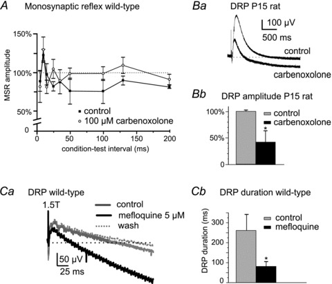
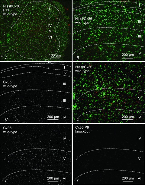
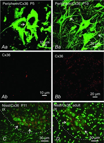
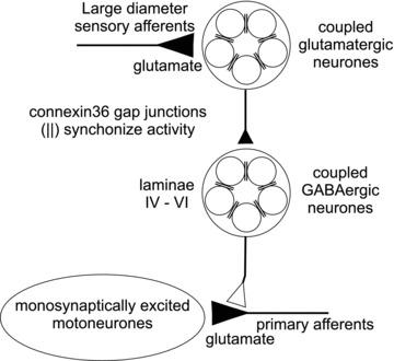
Electrical synapses formed by gap junctions containing connexin36 (Cx36) promote synchronous activity of interneurones in many regions of mammalian brain; however, there is limited information on the role of electrical synapses in spinal neuronal networks. Here we show that Cx36 is widely distributed in the spinal cord and is involved in mechanisms that govern presynaptic inhibition of primary afferent terminals. Electrophysiological recordings were made in spinal cord preparations from 8- to 11-day-old wild-type and Cx36 knockout mice. Several features associated with presynaptic inhibition evoked by conditioning stimulation of low threshold hindlimb afferents were substantially compromised in Cx36 knockout mice. Dorsal root potentials (DRPs) evoked by low intensity stimulation of sensory afferents were reduced in amplitude by 79% and in duration by 67% in Cx36 knockouts. DRPs were similarly affected in wild-types by bath application of gap junction blockers. Consistent with presynaptic inhibition of group Ia muscle spindle afferent terminals on motoneurones described in adult cats, conditioning stimulation of an adjacent dorsal root evoked a long duration inhibition of monosynaptic reflexes recorded from the ventral root in wild-type mice, and this inhibition was antagonized by bicuculline. The same conditioning stimulation failed to inhibit monosynaptic reflexes in Cx36 knockout mice. Immunofluorescence labelling for Cx36 was found throughout the dorsal and ventral horns of the spinal cord of juvenile mice and persisted in mature animals. In deep dorsal horn laminae, where interneurones involved in presynaptic inhibition of large diameter muscle afferents are located, cells were extensively dye-coupled following intracellular neurobiotin injection. Coupled cells displayed Cx36-positive puncta along their processes. Our results indicate that gap junctions formed by Cx36 in spinal cord are required for maintenance of presynaptic inhibition, including the regulation of transmission from Ia muscle spindle afferents. In addition to a role in presynaptic inhibition in juvenile animals, the persistence of Cx36 expression among spinal neuronal populations in the adult mouse suggests that the contribution of electrical synapses to integrative processes in fully mature spinal cord may be as diverse as that found in other areas of the CNS.


Figures

Figure 1
Figure 1. Experimental setup and comparison of CDP, DRP and MSR in P8–11 wild-type and Cx36 knockout mice
Aa, schematic diagram of set-up for evoking the cord dorsum potential (CDP) and dorsal root potential (DRP). B, CDP recorded from wild-type (Ba) and knockout mice (Bb), evoked by stimulation of the L2 dorsal root at 2.5 and 5 times the intensity of threshold for the most excitable afferents (T) using a rectangular current pulse 0.2 ms in duration. Arrows indicate the longer latency components seen in wild-type mice, but which are absent in Cx36 knockout mice. C, DRP recorded in L3 from a P10 wild-type mouse (grey trace) following single shock stimulation at 1.5T (Ca) and 5T (Cb) of the L2 dorsal root overlaid on the DRP from a Cx36 knockout mouse (Ca and Cb, black trace). The DRP is attenuated and short lasting in mice lacking Cx36. Da, schematic diagram of set-up for evoking monosynaptic reflexes (MSRs) in L3 and for stimulation of the L2 dorsal root for evaluating the effects of conditioning stimulation on the monosynaptic reflex. Db, reflexes recorded from the L3 ventral root evoked by stimulation of the L3 dorsal root. Grey traces, averaged responses from a wild-type mouse; black traces, from a Cx36 knockout mouse. Arrows indicate the point of MSR amplitude measurement. In this and following figures, stimulus artefacts are truncated for illustration purposes.
Figure 8
Figure 8. Immunofluorescence labelling of Cx36 in lumbar spinal cord of adult mouse
A, low magnification of deep dorsal horn and the whole of the ventral horn grey matter (outlined by dotted line). Cx36-puncta are distributed throughout, but are most dense in lamina IX (arrows) and within the boxed area straddling the deep dorsal horn and intermediate zone. B, magnification of a region corresponding to that of the box in A, showing dispersed and clustered Cx36-puncta (arrow). C–E, fluorescence Nissl counterstained (green) sections showing examples of Cx36-puncta (red) densely distributed around relatively small neurones (arrows) located in lamina VI (C), lamina VII (D) and in a mid-region of lamina VIII (E).
Figure 2
Figure 2. Effects of the GABAA antagonist bicuculline on DRPs
A and B, left, typical effects of bath application of 20 μm bicuculline on DRPs recorded in the fourth lumbar segment and evoked by 1.5T stimulation of the L3 dorsal root in wild-type mice (A) and Cx36 knockout mice (B). Right, bar graphs showing averaged effects of bicuculline on DRP amplitude from four experiments, with L3 dorsal root stimulation at 1.5T and 2.5T in wild-type mice (A) and Cx36 knockout mice (B). Results are expressed as means ± SEM; *P < 0.05. The peak amplitudes of DRPs evoked by 1.5T and 2.5T stimulation were attenuated by bicuculline in wild-types (A, bar graphs), whereas the DRPs evoked by 1.5T stimulation were unaffected and those evoked by 2.5T stimulation were increased in knockouts (B).
Figure 3
Figure 3. Effects of conditioning stimulation of the MSR in juvenile wild-type and Cx36 knockout mice
Using the paradigm outlined in Fig. 1Da, effects of a conditioning stimulus to the L2 dorsal root were examined on the MSR evoked by L3 dorsal root stimulation and recorded in the L3 ventral root. Normalized MSR amplitude is on the ordinate and intervals between the conditioning and test stimuli is on the abscissa. MSR amplitude is normalized to the amplitude of the unconditioned reflex. A, mean MSR amplitude (±SEM) following conditioning stimulation from 6 wild-type mice (filled squares). Conditioning stimulation of the L2 dorsal root in wild-types inhibits the L3-evoked MSR for more than 200 ms. Results obtained in adult cat (Eccles et al. 1963) are shown for comparison (open circles). B, bath administration of bicuculline in wild-type mice (n = 4) abolishes long-lasting inhibition of the MSR, showing GABAergic mediation of this inhibition. In the presence of bicuculline, values at conditioning–test intervals greater than 50 ms were significantly different from controls (P < 0.05). C, conditioning stimulation of the L2 dorsal root failed to inhibit the L3-evoked MSR in Cx36 knockout mice (n = 7). Results in wild-types from A plotted for comparison.
Figure 4
Figure 4. Primary afferent depolarization (PAD) in lumbar spinal cord of P10 wild-type and Cx36 knockout mice
A, antidromic activation of afferent fibres recorded in the L3 dorsal root following intraspinal stimulation (test, black arrow) in the dorsal horn of a wild-type mouse (upper trace). A preceding conditioning stimulus (open arrow) to the L2 dorsal root at 1.5T (lower trace) increased the amplitude of the intraspinally evoked antidromic signal, i.e. PAD. B, similar recording from a Cx36 knockout mouse shows the unconditioned antidromic activation of fibres in the dorsal root (upper trace). Conditioning stimulation decreased and often inhibited the antidromic response in knockout mice (lower trace). C, comparison of the effects of conditioning stimulation in wild-type versus Cx36 knockout mice (P < 0.05; n = 6). Values of the antidromic response >100% reflect PAD-evoked increased excitability of the afferents produced by the conditioning stimulation. Stars indicate means significantly different (P < 0.05) from 100% control.
Figure 5
Figure 5. Gap junction blockers reduce presynaptic inhibition of the MSR and DRP amplitude in wild-type mice
A, plot showing prolonged inhibition of the L3 MSR produced by L2 stimulation at conditioning–test intervals >50 ms in P9 wild-type mice (filled symbols) and abolition of this long-lasting MSR inhibition by carbenoxolone (open symbols, n = 3). Ba, example of the depressive actions of carbenoxolone on a DRP recorded in L4 and evoked by 2T stimulation of the third lumbar dorsal root in a P15 rat. Bb, summary of effects of carbenoxolone on DRP amplitude in P15 rats (n = 7). Ca, depressant action of the gap junction blocker mefloquine (5 μm) on a DRP evoked at 1.5T in a P9 wild-type mouse (grey trace, control; black trace, mefloquine). DRP amplitude returned to control levels after washout of mefloquine (dotted trace). Cb, summary of depressant actions of mefloquine on the DRP duration evoked at 1.5T in P9 wild-type mice (n = 6). *P < 0.05.
Figure 6
Figure 6. Immunofluorescence labelling for Cx36 in transverse sections of mouse L4 spinal cord in juvenile mice
Panels A–E are from P11 mice and panel F is from a P9 mouse at the fourth lumbar segment. A, low magnification, fluorescence Nissl counterstained section (green) showing distribution of Cx36-positive puncta (red) in dorsal and ventral grey matter (outlined by dotted line). B and C, magnification of superficial dorsal horn laminae (outlined by dotted lines) with (B) and without (C) Nissl counterstain, showing sparse labelling for Cx36 (red) in lamina I and in outer (IIo) and inner (IIi) lamina II, and moderate labelling in lamina III. D and E, magnification of deeper dorsal horn laminae (outlined by dotted lines) with (D) and without (E) Nissl counterstain, showing abundant Cx36-puncta in laminae IV, V and a portion of VI. F, section from the dorsal horn of a P9 Cx36 knockout mouse showing absence of labelling for Cx36 in a field similar to that shown in E.
Figure 7
Figure 7. Immunofluorescence labelling of Cx36 in mouse spinal cord ventral horn at different developmental stages
Aa and Ba, laser scanning confocal overlay images showing double immunofluorescence labelling for Cx36 (red) and the motoneurone marker peripherin (green) at P5 (Aa) and P10 (Ba). Ab and Bb show only the Cx36 labelling (red) from images in Aa and Ba, respectively. At P5 motoneurones display Cx36-puncta at points of somal contact. At P10, Cx36-puncta are on somata and initial dendrites of motoneurones (Ba, arrows). C, Nissl counterstained (green) sections showing dense labelling for Cx36 (red) in lamina IX at P11 among a cluster of neurones in the ventrolateral region of lamina IX (arrows), and persistence of these puncta in adult (D, arrows). Border between ventrolateral grey and white matter is shown by dotted line.
Figure 9
Figure 9. Immunofluorescence labelling of Cx36 (red) associated with neurobiotin-coupled neurones (green) in deep dorsal horn and intermediate zone of mice at P11
A and B, low magnification showing a neurobiotin-injected neuron (A, arrowhead), giving rise to surrounding neurobiotin-positive neurons (A, arrows), and the same field (B) showing dense immunolabelling for Cx36 in lamina VI. C and D, neurobiotin-injected neuron in lamina V (C, arrowhead), and higher magnification from an adjacent section showing clusters of neurobiotin-positive neurones (D, arrow). E and F, confocal laser scanning images showing overlays of neurobiotin-positive neurones and immunofluorescence labelling for Cx36, with green/red overlap seen as yellow. The image in E is a magnification of the boxed area shown in A. Cx36-puncta are seen associated with dendrites and soma of a neurobiotin-injected neurone (E, arrowhead) and neurobiotin-coupled neurone (F, arrows).
Figure 10
Figure 10. Proposed role of neuronal gap junctions in facilitating presynaptic inhibition
Large diameter sensory afferents contact populations of lumbar spinal interneurones that are excitatory to other interneurones located in intermediate spinal laminae. These second order interneurones release GABA onto the presynaptic terminals of primary afferents causing PAD and a subsequent reduction in glutamate release from sensory afferents. In the circuit depicted here, this presynaptic inhibition results in smaller monosynaptic EPSPs generated in motoneurones by Ia muscle spindle afferents. Gap junctions formed by Cx36 (∥) are hypothesized to couple and synchronize activity in interneurone populations responsible for presynaptic inhibition. Ablation of Cx36 or pharmacological inhibition of gap junctions in wild-types results in a severe impairment of the presynaptic regulation of transmission from large diameter sensory afferents.

References

    1. Allison DW, Wilcox RS, Ellefsen KL, Askew CE, Hansen DM, Wilcox JD, Sandoval SS, Eggett DL, Yanagawa Y, Steffensen SC. Mefloquine effects on ventral tegmental area dopamine and GABA neuron inhibition: a physiologic role for connexin-36 gap junctions. Synapse. 2011;65:805–813. - PMC - PubMed
    1. Bagust J, Green KA, Kerkut GA. Strychnine sensitive inhibition in the dorsal horn of mammalian spinal cord. Brain Res. 1981;217:425–429. - PubMed
    1. Bagust J, Forsythe D, Kerkut GA. Demonstration of the synaptic origin of primary afferent depolarisation (PAD) in the isolated spinal cord of the hamster. Brain Res. 1985;341:385–389. - PubMed
    1. Bennett MVL. Gap junctions as electrical synapses. J Neurocytol. 1997;26:349–366. - PubMed
    1. Bennett MV, Zukin RS. Electrical coupling and neuronal synchronization in the mammalian brain. Neuron. 2004;41:495–511. - PubMed

Publication types

LinkOut - more resources