

The role of the paraventricular nucleus of the hypothalamus in the regulation of cardiac and renal sympathetic nerve activity in conscious normal and heart failure sheep
- PMID: 22615431
- PMCID: PMC3630774
- DOI: 10.1113/jphysiol.2012.236059
The role of the paraventricular nucleus of the hypothalamus in the regulation of cardiac and renal sympathetic nerve activity in conscious normal and heart failure sheep
Abstract
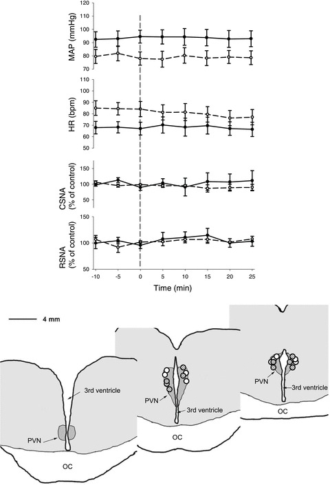
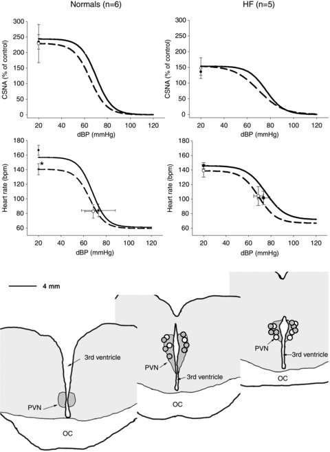
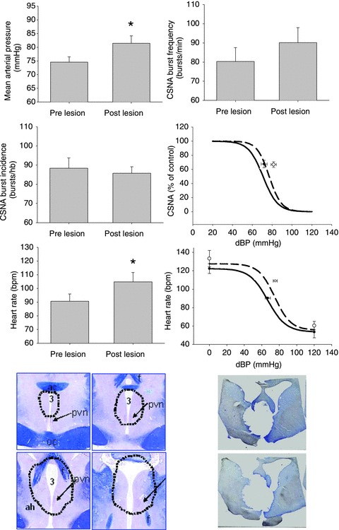
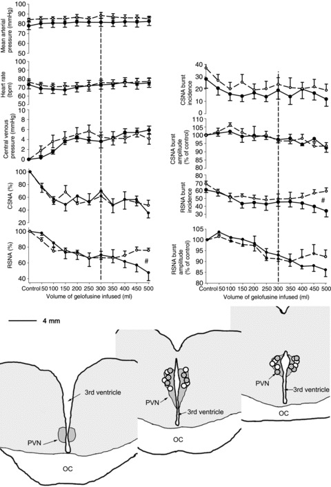
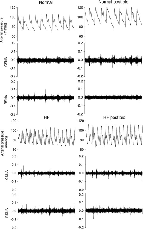
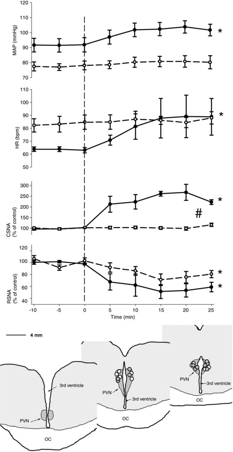
The paraventricular nucleus of the hypothalamus (PVN) plays a major role in central cardiovascular and volume control, and has been implicated in controlling sympathetic nerve activity (SNA) during volume expansion and in heart failure (HF). The objectives were to determine the role of the PVN on cardiac and renal SNA (CSNA and RSNA) in conscious normal sheep and sheep with pacing-induced heart failure. In normovolaemic sheep in the normal state and in HF, bilateral microinjection of the GABA agonist muscimol (2 mm, 500 nl), had no effects on resting mean arterial pressure (MAP), heart rate (HR), CSNA or RSNA. In addition, neither chemical inhibition of the PVN using the inhibitory amino acid glycine (0.5 m, 500 nl), nor electrolytic lesion of the PVN reduced the elevated level of CSNA in HF. Dysinhibition of the PVN with bilateral microinjection of bicuculline (1 mm, 500 nl) in normal sheep increased MAP, HR and CSNA, but decreased RSNA, whereas in HF bicuculline had no effects on MAP, HR or CSNA, but inhibited RSNA. During volume expansion in normal sheep, muscimol reversed the inhibition of RSNA, but not of CSNA. In summary, removal of endogenous GABAergic inhibition to the PVN indicated that CSNA is normally under inhibitory control. Although this inhibition was absent in HF, the responses to pharmacological inhibition, or lesion of the PVN, indicates that it does not drive the increased CSNA in HF. These findings indicate the PVN has a greater influence on RSNA than CSNA in the resting state in normal and HF sheep, and during volume expansion in normal sheep.
Figures
Comment in
-
The hypothalamic paraventricular nucleus may not be at the heart of sympathetic outflow.J Physiol. 2013 Jan 1;591(1):1. doi: 10.1113/jphysiol.2012.237065. J Physiol. 2013. PMID: 23281481 Free PMC article. No abstract available.
Similar articles
-
Central exogenous nitric oxide decreases cardiac sympathetic drive and improves baroreflex control of heart rate in ovine heart failure.Am J Physiol Regul Integr Comp Physiol. 2014 Aug 1;307(3):R271-80. doi: 10.1152/ajpregu.00057.2014. Epub 2014 May 21. Am J Physiol Regul Integr Comp Physiol. 2014. PMID: 24848361
-
Cardiac sympathoexcitation in heart failure.Auton Neurosci. 2013 Apr;175(1-2):76-84. doi: 10.1016/j.autneu.2012.10.017. Epub 2013 Jan 14. Auton Neurosci. 2013. PMID: 23332860 Review.
-
Changes in GABAergic inputs in the paraventricular nucleus maintain sympathetic vasomotor tone in chronic heart failure.Auton Neurosci. 2012 Nov 2;171(1-2):41-8. doi: 10.1016/j.autneu.2012.10.005. Epub 2012 Nov 10. Auton Neurosci. 2012. PMID: 23146621
-
Reduced endogenous GABA-mediated inhibition in the PVN on renal nerve discharge in rats with heart failure.Am J Physiol Regul Integr Comp Physiol. 2002 Apr;282(4):R1006-15. doi: 10.1152/ajpregu.00241.2001. Am J Physiol Regul Integr Comp Physiol. 2002. PMID: 11893604
-
Specific control of sympathetic nerve activity to the mammalian heart and kidney.Exp Physiol. 2010 Jan;95(1):34-40. doi: 10.1113/expphysiol.2008.046342. Epub 2009 Jul 17. Exp Physiol. 2010. PMID: 19617268 Review.
Cited by
-
NK1-receptor-expressing paraventricular nucleus neurones modulate daily variation in heart rate and stress-induced changes in heart rate variability.Physiol Rep. 2014 Dec 3;2(12):e12207. doi: 10.14814/phy2.12207. Print 2014 Dec 1. Physiol Rep. 2014. PMID: 25472606 Free PMC article.
-
Hypothalamic Corticotropin-Releasing Hormone Contributes to Hypertension in Spontaneously Hypertensive Rats.J Neurosci. 2023 Jun 14;43(24):4513-4524. doi: 10.1523/JNEUROSCI.2343-22.2023. Epub 2023 May 9. J Neurosci. 2023. PMID: 37160364 Free PMC article.
-
Howard Florey and neuroscience.J Physiol. 2013 Jan 1;591(1):33-4. doi: 10.1113/jphysiol.2012.248963. J Physiol. 2013. PMID: 23281485 Free PMC article. No abstract available.
-
The negative effects of social bond disruption are partially ameliorated by sertraline administration in prairie voles.Auton Neurosci. 2019 Jul;219:5-18. doi: 10.1016/j.autneu.2019.03.003. Epub 2019 Mar 15. Auton Neurosci. 2019. PMID: 31122602 Free PMC article.
-
Renal Afferents.Curr Hypertens Rep. 2016 Sep;18(9):69. doi: 10.1007/s11906-016-0676-z. Curr Hypertens Rep. 2016. PMID: 27595156 Free PMC article. Review.
References
-
- Akama H, McGrath BP, Badoer E. Volume expansion fails to normally activate neural pathways in the brain of conscious rabbits with heart failure. J Auton Nerv Sys. 1998;73:54–62. - PubMed
-
- Akine A, Montanaro M, Allen AM. Hypothalamic paraventricular nucleus inhibition decreases renal sympathetic nerve activity in hypertensive and normotensive rats. Auton Neurosci. 2003;108:17–21. - PubMed
-
- Badoer E. Hypothalamic paraventricular nucleus and cardiovascular regulation. Clin Exp Pharmacol Physiol. 2001;28:95–99. - PubMed
-
- Badoer E, Ng CW, De Matteo R. Tonic sympathoinhibition arising from the hypothalamic PVN in the conscious rabbit. Brain Res. 2002;947:17–24. - PubMed
-
- Cano G, Card JP, Sved AF. Dual viral transneuronal tracing of central autonomic circuits involved in the innervation of the two kidneys in rat. J Comp Neurol. 2004;471:462–481. - PubMed
Publication types
MeSH terms
Substances
LinkOut - more resources
Full Text Sources
Medical
Research Materials
Miscellaneous