

A retrospective analysis of histopathology of 64 cases of lepra reactions
- PMID: 22615507
- PMCID: PMC3352632
- DOI: 10.4103/0019-5154.94278
A retrospective analysis of histopathology of 64 cases of lepra reactions
Abstract
Background: Lepra reactions are not always diagnosable under the microscope. We analyzed skin histopathology in 64 cases of lepra reaction.
Aim: To make detailed observations on histopathologic features of type 1 and type 2 lepra reaction (erythema nodosum leprosum, ENL).
Materials and methods: In this retrospective study, we included 64 patients diagnosed during a 3-year period as lepra reaction based on clinico-pathological co-relation.
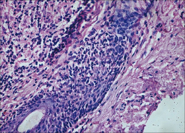
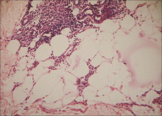
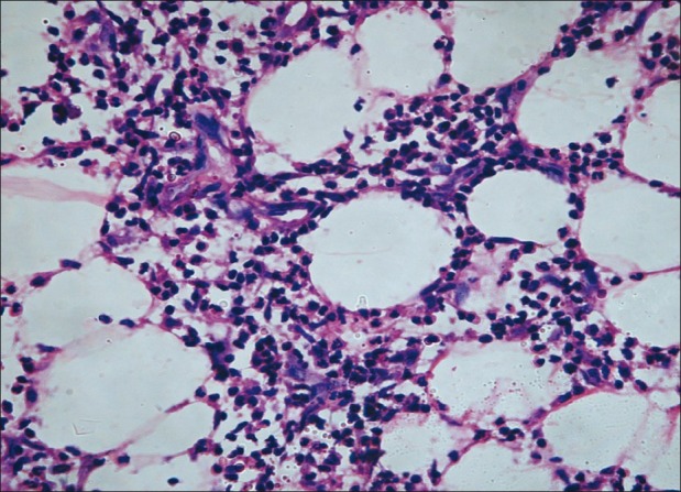
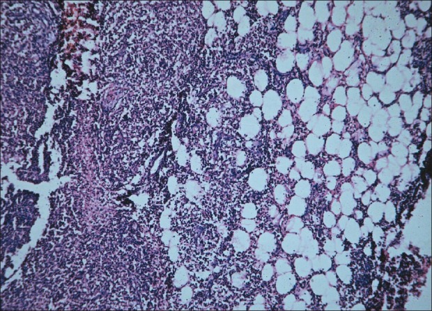
Results: Out of the 64 patients, 22 were of type 1 reaction and 42 of ENL. The most consistent finding in type 1 reaction was papillary dermal edema (86%) followed by pyknosis of lymphocytes (77%) and intercellular edema within granuloma (73%). Surprisingly, folliculotropism of lymphocytes was seen in 55% and subcutaneous infiltration in 36%. In ENL, the most common finding was presence of neutrophils within the granuloma (100%), followed by leukocytoclasia (81%), papillary dermal edema (81%), and neutrophilic panniculitis (69%). Fibrin in the vessel wall or/and granulomas was noted in only 38% while fibrin thrombi in the vessel walls were seen in only 12% of cases.
Conclusion: Infiltration of macrophage granulomas by neutrophils is a reliable sign of ENL. Classical signs of vasculitis are not always present in ENL. Folliculotropism and lymphocytic panniculitis were frequent in type 1 reactions while neutrophilic panniculitis was common with ENL.
Keywords: Erythema nodosum leprosum; lepra reaction; vasculitis.
Conflict of interest statement
Figures
References
-
- Lucas S. Bacterial diseases. In: David E, Rosalie E, Bernett J, George M, editors. Lever's histopathology of the skin. 9th ed. Philadelphia: Lippincott Williams and Wilkins; 2005. p. 575.
-
- Hussain R, Lucas SB, Kifayet A, Jamil S, Raynes J, Uqaili Z, et al. Clinical and histological discrepancies in diagnosis of ENL reactions classified by assessment of acute phase proteins SAA and CRP. Int J Lepr Other Mycobact Dis. 1995;63:222–30. - PubMed
-
- Sehgal VN, Gautam RK, Koranne RV, Beohar PC. The histopathological study of type 1(Lepra) and type 2 (ENL) reactions in leprosy. Indian J Lepr. 1986;58:240–3. - PubMed
-
- Lazaro-Medina A, Tianco EA, Avila JM. Additional markers for the type I reactional states of borderline leprosy. Infect Immun. 1983;39:388–93. - PubMed
-
- Lockwood DN, Lucas SB, Desikan KV, Ebenezer G, Suneetha S, Nicholls P. The histological diagnosis of leprosy type 1 reactions: Identification of key variables and an analysis of the process of histological diagnosis. J Clin Pathol. 2008;61:595–600. - PubMed
LinkOut - more resources
Full Text Sources