. 2012;8(5):e1002705.
doi: 10.1371/journal.ppat.1002705. Epub 2012 May 17.

Immunogenetic mechanisms driving norovirus GII.4 antigenic variation

Affiliations

Immunogenetic mechanisms driving norovirus GII.4 antigenic variation

Lisa C Lindesmith et al. PLoS Pathog. 2012.

Abstract

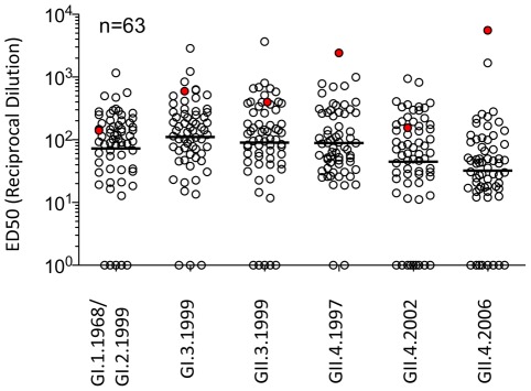
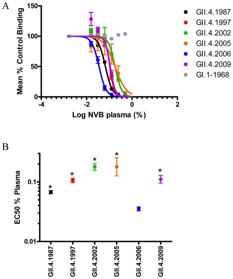
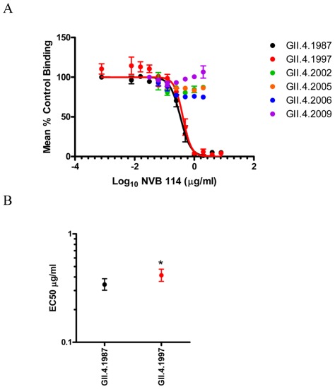
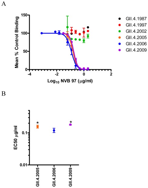
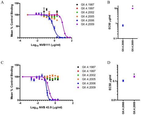
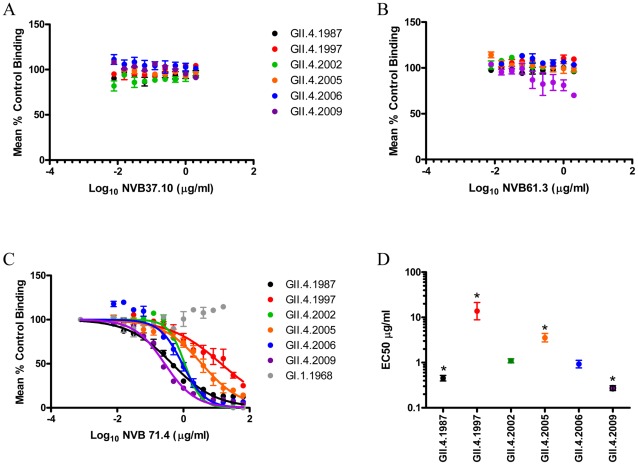
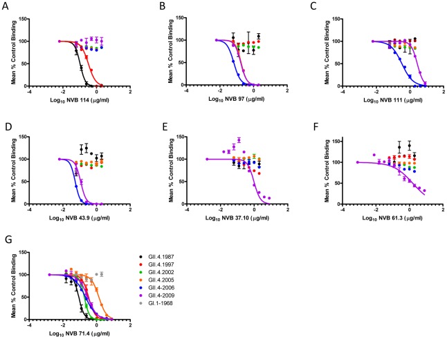
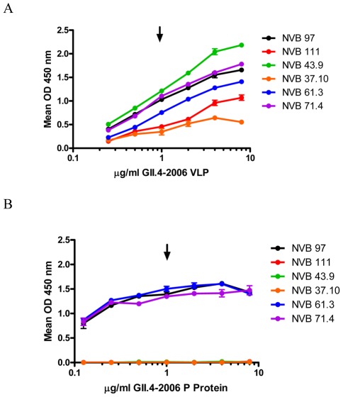
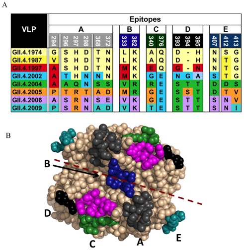
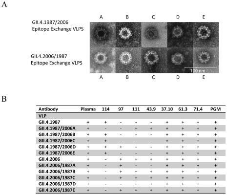
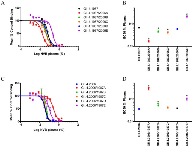
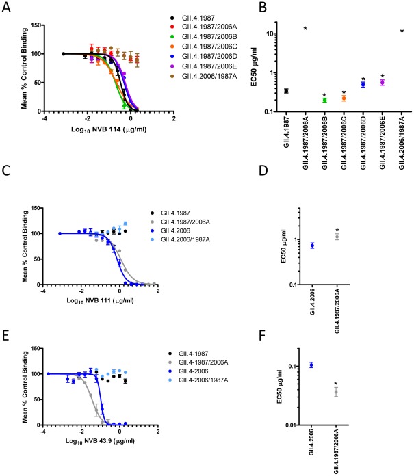
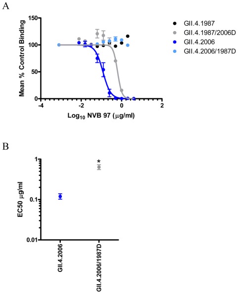
Noroviruses are the principal cause of epidemic gastroenteritis worldwide with GII.4 strains accounting for 80% of infections. The major capsid protein of GII.4 strains is evolving rapidly, resulting in new epidemic strains with altered antigenic potentials. To test if antigenic drift may contribute to GII.4 persistence, human memory B cells were immortalized and the resulting human monoclonal antibodies (mAbs) characterized for reactivity to a panel of time-ordered GII.4 virus-like particles (VLPs). Reflecting the complex exposure history of the volunteer, human anti-GII.4 mAbs grouped into three VLP reactivity patterns; ancestral (1987-1997), contemporary (2004-2009), and broad (1987-2009). NVB 114 reacted exclusively to the earliest GII.4 VLPs by EIA and blockade. NVB 97 specifically bound and blocked only contemporary GII.4 VLPs, while NBV 111 and 43.9 exclusively reacted with and blocked variants of the GII.4.2006 Minerva strain. Three mAbs had broad GII.4 reactivity. Two, NVB 37.10 and 61.3, also detected other genogroup II VLPs by EIA but did not block any VLP interactions with carbohydrate ligands. NVB 71.4 cross-neutralized the panel of time-ordered GII.4 VLPs, as measured by VLP-carbohydrate blockade assays. Using mutant VLPs designed to alter predicted antigenic epitopes, two evolving, GII.4-specific, blockade epitopes were mapped. Amino acids 294-298 and 368-372 were required for binding NVB 114, 111 and 43.9 mAbs. Amino acids 393-395 were essential for binding NVB 97, supporting earlier correlations between antibody blockade escape and carbohydrate binding variation. These data inform VLP vaccine design, provide a strategy for expanding the cross-blockade potential of chimeric VLP vaccines, and identify an antibody with broadly neutralizing therapeutic potential for the treatment of human disease. Moreover, these data support the hypothesis that GII.4 norovirus evolution is heavily influenced by antigenic variation of neutralizing epitopes and consequently, antibody-driven receptor switching; thus, protective herd immunity is a driving force in norovirus molecular evolution.


Conflict of interest statement

The authors have declared that no competing interests exist.

Figures

Figure 1
Figure 1. EIA Reactivity of plasma collected from healthy donors against norovirus VLPs.
VLP-specific IgG titers in 63 plasma samples collected in early 2009 were measured by EIA using a panel of norovirus VLPs as antigen. Reciprocal ED50 dilutions (see Materials and Methods) are shown. Highlighted in red is the plasma sample of the donor NVB selected for the isolation of human mAbs. ED50 values below 10 were scored as negative and assigned a value of 1. The line shows the geometric mean value.
Figure 2
Figure 2. Characterization of donor NVB plasma blockade of norovirus VLPs.
Panel A: PGM binding blockade activity. Sigmoidal curves were fit to the mean percent control binding calculated by comparing the amount of VLP bound to PGM in the presence of antibody pretreatment to the amount of VLP bound in the absence of antibody pretreatment. Error bars represent the standard error of the mean. Panel B: Mean EC50 (% plasma) for blockade of each VLP. * VLPs with EC50 values significantly different from the EC50 for GII.4.2006.
Figure 3
Figure 3. Human mAb NVB 114 recognizes a blockade epitope restricted to early GII.4 strains.
Panel A: PGM binding blockade activity. Sigmoidal curves were fit to the mean percent control binding calculated by comparing the amount of VLP bound to PGM in the presence of antibody pretreatment to the amount of VLP bound in the absence of antibody pretreatment. Error bars represent the standard error of the mean. Panel B: Mean EC50 (µg/ml) for blockade of each VLP. * VLP with EC50 value significantly different from the EC50 for GII.4.1987.
Figure 4
Figure 4. Human mAb NVB 97 recognizes a blockade epitope restricted to contemporary GII.4 strains.
Panel A: PGM binding blockade activity. Sigmoidal curves were fit to the mean percent control binding calculated by comparing the amount of VLP bound to PGM in the presence of antibody pretreatment to the amount of VLP bound in the absence of antibody pretreatment. Error bars represent the standard error of the mean. Panel B: Mean EC50 (µg/ml) for blockade of each VLP. * VLP with EC50 value significantly different from the EC50 for GII.4.2006.
Figure 5
Figure 5. Human mAbs NVB 111 and 43.9 recognize a blockade epitope restricted to Minerva variants.
Panel A (NVB 111) and C (NVB 43.9): PGM binding blockade activity. Sigmoidal curves were fit to the mean percent control binding calculated by comparing the amount of VLP bound to PGM in the presence of antibody pretreatment to the amount of VLP bound in the absence of antibody pretreatment. Error bars represent the standard error of the mean. Panels B (NVB 111) and D (NVB 43.9): Mean EC50 (µg/ml) for blockade of each VLP. * VLP with EC50 value significantly different from the EC50 for GII.4.2006.
Figure 6
Figure 6. Human mAbs NVB 37.10, 61.3 and 71.4 recognize a conserved epitope.
PGM binding blockade activity of NVB 37.10 (Panel A), NVB 61.3 (Panel B) and NVB 71.4 (Panel C). Sigmoidal curves were fit to the mean percent control binding calculated by comparing the amount of VLP bound to PGM in the presence of antibody pretreatment to the amount of VLP bound in the absence of antibody pretreatment. Error bars represent the standard error of the mean. Panel D: Mean EC50 (µg/ml) for blockade of each VLP by NVB 71.4. * VLPs with EC50 value significantly different from the EC50 for GII.4.2006.
Figure 7
Figure 7. Blockade of Bi-HBGAs by Hu mAbs.
Bi-HBGA binding blockade activity of NVB 114 (Panel A), NVB 97 (Panel B), NVB 111 (Panel C), NBV 43.9 (Panel D), NVB 37.10 (Panel E), NVB 61.3 (Panel F) and NVB 71.4 (Panel G). Sigmoidal curves were fit to the mean percent control binding calculated by comparing the amount of VLP bound to Bi-HBGA in the presence of antibody pretreatment to the amount of VLP bound in the absence of antibody pretreatment. Error bars represent the standard error of the mean. EC50 values are reported in Table S2.
Figure 8
Figure 8. EIA Reactivity of mAbs to GII.4.2006 VLPs and P proteins.
Increasing concentrations of GII.4.2006 VLP (Panel A) or P protein (Panel B) were assayed for reactivity to the hu mAbs by EIA. Arrow indicates 1 µg/ml, the concentration of GII.4.2006 VLP scored as postive by EIA (Figure S2 and Table 2). Bars are SEM.
Figure 9
Figure 9. Predicted GII.4 norovirus evolving blockade epitopes.
Bioinformatic approaches predicted five antibody epitopes on the surface of GII.4 noroviruses that appeared to be evolving over time and to correlate with the emergence of new GII.4 outbreak strains. Panel A: Amino acid variation of Epitopes A–E by GII.4 strain. Panel B: Predicted epitopes were expanded to include interacting amino acids within 8A. Epitope A (grey), Epitope B (blue), Epitope C (green), Epitope D (black), Epitope E (teal) and HBGA binding sites (magenta) mapped onto the P domain dimer of GII.4.2002.
Figure 10
Figure 10. Characterization of Epitope A through E exchanged VLPs.
Panel A: Electron microscopy visualization of negative-stained epitope-exchanged VLPs. Panel B: Antibody and PGM binding phenotypes of epitope-exchanged VLPs.
Figure 11
Figure 11. Characterization of donor NVB plasma reactivity to engineered epitope-exchanged VLPs.
PGM binding blockade activity against GII.4.1987 (Panel A) and GII.4.2006 (Panel C) Epitope A–E chimeric VLPs. Sigmoidal curves were fit to the mean percent control binding calculated by comparing the amount of VLP bound to PGM in the presence of antibody pretreatment to the amount of VLP bound in the absence of antibody pretreatment. Error bars represent the standard error of the mean. Mean EC50 (% plasma) for blockade of each GII.4.1987 (Panel B) and GII.4.2006 (Panel D) epitope-exchanged VLP. * VLPs with EC50 values significantly different from the EC50 for each parental VLP.
Figure 12
Figure 12. Epitope A comprises an evolving GII.4 blockade epitope recognized by NVB 114, 111 and 43.9.
Hu mAb PGM binding blockade activity against GII.4.1987 epitope-exchanged VLPs for NVB 114 (Panel A) and Epitope A exchanged between GII.4.1987 and GII.4.2006 (Panels C and E). Sigmoidal curves were fit to the mean percent control binding calculated by comparing the amount of VLP bound to PGM in the presence of antibody pretreatment to the amount of VLP bound in the absence of antibody pretreatment. Error bars represent the standard error of the mean. Mean EC50 for blockade of each GII.4.1987 exchange epitope (Panel B) and Epitope A exchange VLPs (Panel D and F). * VLPs with EC50 values significantly different from the EC50 for each parental VLP.
Figure 13
Figure 13. Amino acids 393–395 comprise a blockade epitope for contemporary GII.4 strains recognized by NVB 97.
Panel A: NVB 97 PGM binding blockade activity against Epitope D exchange VLPs. Sigmoidal curves were fit to the mean percent control binding calculated by comparing the amount of VLP bound to PGM in the presence of antibody pretreatment to the amount of VLP bound in the absence of antibody pretreatment. Error bars represent the standard error of the mean. Panel B: Mean EC50 for blockade of each blocked VLP. * indicates VLPs with significantly different EC50 compared GII.4.2006.
Figure 14
Figure 14. Summary of exchange mutant VLPs containing blockade Epitopes A and D and the reactivity pattern of the human mAbs that recognize these epitopes.

References

    1. Updated norovirus outbreak management and disease prevention guidelines. MMWR Recomm Rep. 2011;60:1–18. - PubMed
    1. Hutson AM, Atmar RL, Estes MK. Norovirus disease: changing epidemiology and host susceptibility factors. Trends Microbiol. 2004;12:279–287. - PMC - PubMed
    1. Estes MK, Prasad BV, Atmar RL. Noroviruses everywhere: has something changed? Curr Opin Infect Dis. 2006;19:467–474. - PubMed
    1. Koopmans M, Vinj inverted question marke J, de Wit M, Leenen I, van der Poel W, et al. Molecular epidemiology of human enteric caliciviruses in The Netherlands. J Infect Dis. 2000;181(Suppl 2):S262–269. - PubMed
    1. Norovirus activity–United States, 2006–2007. MMWR Morb Mortal Wkly Rep. 2007;56:842–846. - PubMed

Publication types

MeSH terms