. 2012;8(5):e1002713.
doi: 10.1371/journal.ppat.1002713. Epub 2012 May 17.

Extracellular superoxide dismutase protects Histoplasma yeast cells from host-derived oxidative stress

Affiliations

Extracellular superoxide dismutase protects Histoplasma yeast cells from host-derived oxidative stress

Brian H Youseff et al. PLoS Pathog. 2012.

Abstract

In order to establish infections within the mammalian host, pathogens must protect themselves against toxic reactive oxygen species produced by phagocytes of the immune system. The fungal pathogen Histoplasma capsulatum infects both neutrophils and macrophages but the mechanisms enabling Histoplasma yeasts to survive in these phagocytes have not been fully elucidated. We show that Histoplasma yeasts produce a superoxide dismutase (Sod3) and direct it to the extracellular environment via N-terminal and C-terminal signals which promote its secretion and association with the yeast cell surface. This localization permits Sod3 to protect yeasts specifically from exogenous superoxide whereas amelioration of endogenous reactive oxygen depends on intracellular dismutases such as Sod1. While infection of resting macrophages by Histoplasma does not stimulate the phagocyte oxidative burst, interaction with polymorphonuclear leukocytes (PMNs) and cytokine-activated macrophages triggers production of reactive oxygen species (ROS). Histoplasma yeasts producing Sod3 survive co-incubation with these phagocytes but yeasts lacking Sod3 are rapidly eliminated through oxidative killing similar to the effect of phagocytes on Candida albicans yeasts. The protection provided by Sod3 against host-derived ROS extends in vivo. Without Sod3, Histoplasma yeasts are attenuated in their ability to establish respiratory infections and are rapidly cleared with the onset of adaptive immunity. The virulence of Sod3-deficient yeasts is restored in murine hosts unable to produce superoxide due to loss of the NADPH-oxidase function. These results demonstrate that phagocyte-produced ROS contributes to the immune response to Histoplasma and that Sod3 facilitates Histoplasma pathogenesis by detoxifying host-derived reactive oxygen thereby enabling Histoplasma survival.


Conflict of interest statement

The authors have declared that no competing interests exist.

Figures
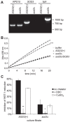
Figure 1
Figure 1. Histoplasma Sod3 encodes an extracellular Cu++-dependent superoxide dismutase.
(A) PCR validation of deletion of the SOD3 gene. Genomic DNA from the SOD3(+) parental strain (WU8) and the sod3Δ strain (OSU13) were tested by PCR for the ribosomal subunit gene RPS15, the wild-type SOD3 gene, and the mutant allele marked with the hygromycin resistance gene (hph). (B) Superoxide dismutase activity in culture filtrates harvested from SOD3(+) (OSU45), sod3Δ (OSU15), and the sod3Δ/SOD3 complemented (OSU49) strains. Detection of superoxide was determined through superoxide-dependent reduction of the WST-1 tetrazolium dye after generation of superoxide using hypoxanthine and xanthine oxidase. Reduction of WST-1 was monitored by absorbance at 438 nm. Buffer or culture filtrates contained 5 µg ovalbumin or total culture filtrate protein, respectively. Asterisks represent significant difference (*** p<0.001) in the inhibition of WST-1 reduction between SOD3(+) and sod3Δ culture filtrates. Data shown is representative of three independent experiments, each performed with triplicate samples. (C) Sod3 activity following Cu++ depletion. Culture filtrates containing 5 µg total protein from SOD3(+) (OSU45) and sod3Δ (OSU15) strains were tested for their ability to inhibit WST-1 reduction by superoxide before (no chelator), after Cu++ depletion (+DDC), and after subsequent repletion with 50 mM Cu++ (+CuSO4). Values represent relative inhibition of WST-1 reduction by culture filtrate samples (n = 3) compared to buffer controls treated in parallel. Asterisks represent significant differences from SOD3(+) culture filtrates (* p<0.05, ** p<0.01).
Figure 2
Figure 2. N-terminal and C-terminal signals direct extracellular localization of Sod3.
(A) Schematic of the Sod3 protein highlighting the predicted signal peptide (SP) and the glycophosphatidyl inositol anchor (GPI) signal motifs. Numbers represent amino acid residues in the Sod3 protein. Shading beneath the Sod3 protein indicates amino acid sequence similarity between G186A, G217B and NAm1 Sod3 proteins ranging from dark (>90% sequence identity) to light (<50% identity). (B) Relative Sod3 activity associated with the yeast cell and soluble extracellular fraction. Superoxide dismutase activities were determined by inhibition of superoxide-dependent WST-1 reduction in the presence of 1×10⁸ yeasts (cell-associated) or the corresponding culture filtrate (soluble) of SOD3(+) (OSU45) and sod3Δ (OSU15) strains (n = 3, each). Inhibition of WST-1 reduction was normalized to reactions in the absence of yeasts or culture filtrate. Asterisks represent significant differences (p<0.001) from SOD3(+) samples. (C) Determination of the localization of GFP when fused to the N-terminus of Sod3. Extracellular or intracellular GFP localization was determined by α-FLAG immunoblot of culture filtrates or cellular lysates from Histoplasma yeast strains expressing FLAG epitope-tagged GFP (GFP:FLAG; OSU88) or GFP with the first 26 amino acids of Sod3 (Sod31–26:GFP:FLAG; OSU102). Cellular lysates were also tested for α-tubulin to demonstrate equal loadings. (D) Localization of Sod3 activity after removal of the C-terminal 26 amino acids. Cell-associated and soluble superoxide dismutase activities of Histoplasma yeasts were determined using 1×10⁸ intact yeasts or their corresponding culture filtrates, respectively. Samples were collected from SOD3(+) (OSU45), sod3Δ (OSU15), and yeasts expressing full length Sod3 (sod3Δ/FLAG:SOD3; OSU116) or Sod3 lacking the putative GPI signal (sod3Δ/FLAG:SOD3 ΔGPI; OSU117). Results were normalized to uninhibited reactions and plotted as the proportion of total inhibitory activity. Asterisks represent significant difference from full length Sod3 (** p<0.01, *** p<0.001). Relative quantitation of Sod3 in culture filtrates was determined by α-FLAG immunoblot and is indicated numbers below.
Figure 3
Figure 3. Histoplasma Sod3 does not alleviate intracellular oxidative stress.
(A) Depletion of intracellular superoxide dismutase activity by SOD1-RNAi but not by loss of Sod3. RNAi-based depletion of Sod1 was done in a GFP-expressing Histoplasma strain (OSU22). GFP fluorescence is shown in colony images of a strain in which gfp was not targeted (gfp(+); OSU103), gfp alone was targeted (gfp-RNAi; OSU104) or two independent isolates in which gfp and SOD1 were co-targeted (gfp:SOD1-RNAi; OSU105). Numbers below the images indicate relative GFP fluorescence. Intracellular superoxide dismutase activity was determined by inhibition of WST-1 reduction using 5 µg of cellular lysate protein from SOD3(+) (OSU45), sod3Δ (OSU15), and the RNAi strains and results plotted relative to uninhibited reactions using 5 µg BSA. Non-significant (ns) and significant (*** p<0.001) differences between SOD3(+) and sod3Δ or between the SOD1(+) strain (gfp(+)) and the gfp-RNAi or SOD1-RNAi strain are indicated above the columns. (B–D) Inhibition of yeast growth by increased intracellular reactive oxygen. Liquid growth of SOD3(+) (OSU45), sod3Δ (OSU15), Sod1-proficient (gfp-RNAi; OSU104), and SOD1-RNAi (gfp:SOD1-RNAi; OSU105) strains was determined by optical density of cultures measured at 595 nm. Intracellular reactive oxygen was increased in yeasts by addition of 5 µM (C) or 10 µM (D) paraquat. Growth curve points represent the mean optical density of replicate cultures (n = 3).
Figure 4
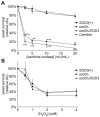
Figure 4. Sod3 protects Histoplasma yeast cells from exogenous superoxide in vitro.
(A) Survival of yeast cells following challenge with superoxide. Yeasts were incubated in increasing amounts of superoxide generated by addition of increasing amounts of xanthine oxidase to hypoxanthine. SOD3(+) (OSU45), sod3Δ (OSU15), sod3Δ/SOD3 (OSU49), and Candida albicans yeasts were incubated for 4 hours at 37°C after which viable colony forming units (cfu) were determined. Results are plotted as relative yeast survival compared to viable cfu of yeasts incubated in the absence of superoxide (0 mU/mL xanthine oxidase). Results represent the mean ± standard deviations from 3 replicate challenges per strain. Asterisks indicate significant differences (** p<0.01, *** p<0.001) from the SOD3(+) strain. (B) Sensitivity of Histoplasma yeasts to hydrogen peroxide. Increasing amounts of hydrogen peroxide were added to Histoplasma yeasts (n = 3 for each strain) at 37°C and the viability of yeasts after 4 hours was determined by enumeration of viable cfu. Results are plotted as relative yeast survival compared to viable cfu of yeasts incubated in the absence of peroxide (0 mM hydrogen peroxide). Data is representative of 3 independent experiments.
Figure 5
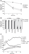
Figure 5. Sod3 protects Histoplasma yeasts from PMN-derived reactive oxygen.
(A) Survival of yeasts after infection of human PMNs. SOD3(+) (OSU45), sod3Δ (OSU15) and Candida albicans yeasts were added to PMNs at a multiplicity of infection (MOI) of 1∶10. Yeast survival was determined by enumeration of viable cfu after 2 and 4 hours of co-incubation of yeasts with PMNs at 37°C. Results are plotted as relative yeast survival (mean ± standard deviation of 3 replicates) compared to viable cfu of yeasts incubated in the absence of PMNs. Significantly decreased survival compared to SOD3(+) yeasts is indicated by asterisks (** p<0.01). (B) Inhibition of yeast killing by PMNs upon inactivation of the NADPH-oxidase. Yeasts were added to PMNs (+ PMNs) and incubated for 4 hours at 37°C and viable cfu were determined. 10 µM diphenylene iodinium (DPI) was added to some assays to inactivate the NADPH-oxidase. Results indicate relative yeast survival (mean ± standard deviation of 3 replicates) compared to viable cfu of yeast incubated in the absence of PMNs (no PMNs). Significant (** p<0.01) or non-significant (ns) reduction in survival compared to yeast in the absence of PMNs is indicated above the respective columns. (C) Reactive oxygen production by PMNs in response to Histoplasma yeasts. Histoplasma yeasts were added to PMNs at an MOI of 1∶1 in the presence of the luminol ROS-detection reagent and the luminol luminescence measured over time. PMNs and yeasts were co-incubated in the presence (open symbols) or absence (closed symbols) of 10 µM DPI to inhibit the NADPH-oxidase. Data points represent the mean luminescence (n = 3).
Figure 6
Figure 6. Sod3 protects Histoplasma yeasts from ROS produced by activated macrophages.
(A–B) Survival of yeasts after infection of resting (A) or cytokine-activated (B) murine macrophages. SOD3(+) (OSU45), sod3Δ (OSU15) and Candida albicans yeasts were added to resident peritoneal macrophages at an MOI of 1∶50. Yeast survival was determined by enumeration of viable cfu after 2 and 4 hours of co-incubation of yeasts with macrophages at 37°C. In (B), 10 U TNFα and 100 U IFNγ were added to macrophages 24 hours prior to infection to enhance ROS production. Results are plotted as relative yeast survival (mean ± standard deviation of 3 replicates) compared to viable cfu of yeasts incubated in the absence of macrophages. Significantly decreased survival compared to SOD3(+) yeasts is indicated by asterisks (* p<0.05, ** p<0.01, *** p<0.001). (C) Prevention of yeast killing by macrophages after inhibition of the NADPH-oxidase. Yeasts were added to resting and to IFNγ/TNFα-activated macrophages and incubated for 4 hours at 37°C in the absence or presence of 10 µM diphenylene iodinium (DPI) and viable cfu were determined. Results indicate relative yeast survival (mean ± standard deviation of 3 replicates) compared to viable cfu of yeasts incubated in the absence of macrophages. Significant (** p<0.01) or non-significant (ns) reduction in survival compared to yeasts in the absence of macrophages is indicated above the respective columns. (D) Reactive oxygen production by activated macrophages in response to Histoplasma yeasts. Histoplasma yeasts were added to resting or activated macrophages at an MOI of 1∶1 in the presence of the luminol ROS-detection reagent and the luminol luminescence measured over time. Macrophages and yeasts were co-incubated in the presence (open symbols) or absence (closed symbols) of 10 µM DPI to inhibit the NADPH-oxidase. Data points represent the mean luminescence (n = 3).
Figure 7
Figure 7. Histoplasma virulence in vivo requires Sod3.
(A) Kinetics of sublethal lung infection by Histoplasma. Wild-type C57BL/6 mice were intranasally infected with approximately 1×10⁴ SOD3(+) (OSU45), sod3Δ (OSU15), or sodΔ/SOD3 (OSU49) Histoplasma yeasts. At 4 day intervals post-infection, the fungal burden in lungs was determined by quantitative platings for Histoplasma cfu. (B) Kinetics of dissemination following lung infection with Histoplasma. At each time point, organs were harvested and the fungal burden in spleen tissue was determined by quantitative platings for cfu. In (A) and (B), each data point represents cfu counts per organ from an individual animal (n = 5 per time point) and horizontal bars represent the mean fungal burden. Asterisks indicate significant differences at each time point from animals infected with SOD3(+) organisms (* p<0.05, ** p<0.01, *** p<0.001). The actual inoculum dose is shown in graphs at day 0. The limit of detection is 100 cfu for lungs and 60 cfu for spleen tissue. (C) Inflammation and pathology of lung tissue following Histoplasma infection. Wild-type C57BL/6 mice were infected with SOD3(+) (OSU45), sod3Δ (OSU15), or sodΔ/SOD3 (OSU49) yeast and at 4 days post-infection, lungs were harvested and sections stained with hematoxylin and eosin. Arrowheads indicate detectable clusters of yeast cells. Scale bars represent 50 µm.
Figure 8
Figure 8. Lethal infection by Histoplasma requires Sod3 function.
Kinetics of mouse survival after infection with a lethal dose of Histoplasma yeasts. Wild-type C57BL/6 mice were intranasally infected with 7×10⁶ SOD3(+) (OSU45), sod3Δ (OSU15), or sodΔ/SOD3 (OSU49) Histoplasma yeasts (n = 8 per strain). Survival time of mice infected with sod3Δ yeasts differs significantly from that of infections with SOD3(+) and sodΔ/SOD3 (p<0.0001).
Figure 9
Figure 9. Sod3 facilitates infection through detoxification of host reactive oxygen.
Kinetics of sublethal lung infection by Histoplasma in animals competent for ROS production (A) or animals lacking the NADPH-oxidase function (B). Mice were intranasally infected with approximately 1×10⁴ SOD3(+) (OSU45) or sod3Δ (OSU15) Histoplasma yeasts. At 2, 4, 8, and 15 days post-infection, the fungal burden in lungs was determined by quantitative platings for Histoplasma cfu. (A) Respiratory infection of Phox(+/+) mice isogenic to the p47phox knock-outs. (B) Respiratory infection of p47phox knock-out (Phox(−/−)) mice. Each data point represents cfu counts per lung from an individual animal (n = 3 per time point) and horizontal bars represent the mean fungal burden. Non-significant (ns) or significant differences (* p<0.05, ** p<0.01) from animals infected with SOD3(+) organisms is indicated above the respective columns. The actual inoculum dose is shown in graphs at day 0. The limit of detection is 100 cfu.

References

    1. Imlay JA. Pathways of oxidative damage. Annu Rev Microbiol. 2003;57:395–418. - PubMed
    1. Johnston RB, Jr, Kitagawa S. Molecular basis for the enhanced respiratory burst of activated macrophages. Fed Proc. 1985;44:2927–2932. - PubMed
    1. Murray HW, Spitalny GL, Nathan CF. Activation of mouse peritoneal macrophages in vitro and in vivo by interferon-gamma. J Immunol. 1985;134:1619–1622. - PubMed
    1. Ajello L. The medical mycological iceberg. HSMHA Health Rep. 1971;86:437–448. - PMC - PubMed
    1. Fleischmann J, Wu-Hsieh B, Howard DH. The intracellular fate of Histoplasma capsulatum in human macrophages is unaffected by recombinant human interferon-gamma. J Infect Dis. 1990;161:143–145. - PubMed

Publication types