. 2012;7(5):e37091.
doi: 10.1371/journal.pone.0037091. Epub 2012 May 15.

Phenotypic and functional changes in blood monocytes following adherence to endothelium

Affiliations

Phenotypic and functional changes in blood monocytes following adherence to endothelium

Colin Tso et al. PLoS One. 2012.

Abstract

Objective: Blood monocytes are known to express endothelial-like genes during co-culture with endothelium. In this study, the time-dependent change in the phenotype pattern of primary blood monocytes after adhering to endothelium is reported using a novel HLA-A2 mistyped co-culture model.

Methods and results: Freshly isolated human PBMCs were co-cultured with human umbilical vein endothelial cells or human coronary arterial endothelial cells of converse human leukocyte antigen A2 (HLA-A2) status. This allows the tracking of the PBMC-derived cells by HLA-A2 expression and assessment of their phenotype pattern over time. PBMCs that adhered to the endothelium at the start of the co-culture were predominantly CD11b+ blood monocytes. After 24 to 72 hours in co-culture, the endothelium-adherent monocytes acquired endothelial-like properties including the expression of endothelial nitric oxide synthase, CD105, CD144 and vascular endothelial growth factor receptor 2. The expression of monocyte/macrophage lineage antigens CD14, CD11b and CD36 were down regulated concomitantly. The adherent monocytes did not express CD115 after 1 day of co-culture. By day 6, the monocyte-derived cells expressed vascular cell adhesion molecule 1 in response to tumour necrosis factor alpha. Up to 10% of the PBMCs adhered to the endothelium. These monocyte-derived cells contributed up to 30% of the co-cultured cell layer and this was dose-dependent on the PBMC seeding density.

Conclusions: Human blood monocytes undergo rapid phenotype change to resemble endothelial cells after adhering to endothelium.


Conflict of interest statement

Competing Interests: The authors have declared that no competing interests exist.

Figures

Figure 1
Figure 1. Endothelial adherence of blood monocytes.
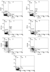
PBMCs were isolated from HLA-A2+ donors and incubated with HLA-A2− HUVECs (1×10⁶ cells/well) for 2 h, after which the non-adherent PBMCs were removed by washing. The co-cultured cell layers were immediately analysed with dual-colour flow cytometry for HLA-A2 and (A) CD34, (B) CD14, (C) CD11b, (D) CD16, (E) CD105 and (F) CD144 expression. Representative plots from 4–6 individual experiments are shown. (G) Two parameters dot plot showing typical isotype controls.
Figure 2
Figure 2. Down-regulation of CD14 and CD11b expression in the endothelium-adherent monocytes during co-culture.
HLA-A2+ PBMCs (1×10⁶ cells/well) were incubated for 2 h (Day 0) with HLA-A2− HUVECs, after which the non-adherent cells were removed by washing. The cell layers were assessed by dual-colour flow cytometry for HLA-A2 and (A) CD14 expression on Day 1 and (B) CD11b expression on Day 3 of co-culture. These are representative plots from 4–7 individual experiments. The reduction in CD14 expression from Day 0 to Day 5 (C) and CD11b expression from Day 0 to Day 3 (D) is also shown.
Figure 3
Figure 3. Increased expression of CD105 and CD144 in the endothelium-adherent monocytes during co-culture.
HLA-A2+ PBMCs (1×10⁶ cells/well) were incubated for 2 h (Day 0) with HLA-A2− HUVECs, after which the non-adherent cells were removed by washing. The cell layers were maintained in co-culture up to Day 6, then assessed by dual-colour flow cytometry for HLA-A2 and (A) CD105 and (B) CD144 expression on Day 3 of co-culture. Representative plots from 4–7 individual experiments are shown. The increase in CD105 from Day 0 to Day 6 (C) and CD144 expression from Day 0 to Day 6 (D) is also shown.
Figure 4
Figure 4. Phenotype change from HLA-A2+/CD11b+/CD105− to HLA-A2+/CD11b−/CD105+ on endothelium-adherent blood monocyte-derived cells with increase in size and granularity during co-culture.
HLA-A2+ PBMCs (1×10⁶ cells/well) were incubated for 2 h (Day 0) with HLA-A2− HUVECs, after which the non-adherent cells were removed by washing. The cell layers were analysed by three-colour flow cytometry staining for HLA-A2, CD11b and CD105 on (A) Day 1 and (B) Day 2. These plots were gated for HLA-A2+ cells. Forward scatter/side scatter dot plots gated for HLA-A2+ cells on Day 0 (C) and Day 2 (D) was shown. These are representative of 2 individual experiments.
Figure 5
Figure 5. Increased expression of VEGFR2 and VCAM-1, reduced expression of CD36 and expression of CD31 and ICAM-1 in the endothelium-adherent monocytes in co-culture.
HLA-A2+ PBMCs (1×10⁶ cells/well) were incubated for 2 h (Day 0) with HLA-A2− HUVEC, after which the non-adherent cells were removed by washing. The cell layers were analysed by dual-colour flow cytometry for HLA-A2 and VEGFR2 expression on Day 1 (A) and Day 2 (B) of co-culture, VCAM-1 expression in the absence of TNF-α on Day 6 of co-culture (C), VCAM-1 expression on Day 6 of co-culture after 24 hours of stimulation with TNF-α (10 ng/ml) (D), CD36 expression on Day 0 (E) and Day 1 (F) of co-culture, as well as CD31 (G) and ICAM-1 (H) expression on Day 0. Representative plots from 2–5 individual experiments are shown.
Figure 6
Figure 6. Expression of eNOS in endothelium-adherent monocytes in co-culture.
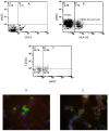
Dual-colour flow cytometry analysis of CD14 and eNOS expression on freshly-isolated PBMCs before co-culture (representative of 2 individual experiments) is shown (A). HLA-A2+ PBMCs (1×10⁶ cells/well) were incubated for 2 h with HLA-A2− HUVECs, after which the non-adherent PBMCs were removed by washing. (B) Shows the flow cytometry analysis of the cell layers for HLA-A2 and eNOS expression on Day 1 of co-culture (representative of 3 individual experiments). (C) Shows the 2 parameters dot plot from 3-colour flow cytometry analysis on Day 1 of co-culture staining for HLA-A2, CD11b and eNOS. This plot is gated for HLA-A2+ cells (representative of 2 individual experiments). Immunofluorescent micrographs (×60 original magnification) showing endothelial-adherent HLA-A2+ cells (green) and eNOS expression (red) on Day 0 (D) and Day 1 (E) of co-culture (representative of 2 individual experiments). The nuclei were stained with DAPI. Scale bar = 20 µm.
Figure 7
Figure 7. Expression of endothelial antigens in endothelium-adherent monocytes in co-culture with HCAECs.
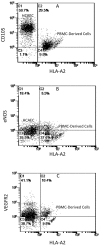
HLA-A2+ PBMCs (1×10⁶ cells/well) were incubated for 2 h (Day 0) with HLA-A2− HCAECs, after which the non-adherent cells were removed by washing. The cell layers were analysed by dual-colour flow cytometry for HLA-A2 and (A) CD105, (B) eNOS and (C) VEGFR2 expression on Day 2 of co-culture.
Figure 8
Figure 8. Quantification of endothelium-adherent monocytes in co-culture.
PBMCs were seeded onto HUVECs of converse HLA-A2 status, co-incubated for 2 h, after which the non-adherent cells were washed off. The percentage of HLA-A2+ cells was evaluated using dual-colour flow cytometry after 24–48 h of co-culture. The graph shows the effect of varying the PBMC seeding densities at 0.25×10⁶, 0.5×10⁶ and 1.0×10⁶ cells/well. The % HLA-A2+ PBMCs co-cultured with the HLA-A2− HUVECs (grey bars) and the % HLA-A2− PBMCs co-cultured with HLA-A2+ HUVECs (black bars) are shown.

Similar articles

Cited by

References

    1. Schubert SY, Benarroch A, Ostvang J, Edelman ER. Regulation of endothelial cell proliferation by primary monocytes. Arteriosclerosis, thrombosis, and vascular biology. 2008;28:97–104. - PubMed
    1. Fernandez Pujol B, Lucibello FC, Gehling UM, Lindemann K, Weidner N, et al. Endothelial-like cells derived from human CD14 positive monocytes. Differentiation. 2000;65:287–300. - PubMed
    1. Schmeisser A, Garlichs CD, Zhang H, Eskafi S, Graffy C, et al. Monocytes coexpress endothelial and macrophagocytic lineage markers and form cord-like structures in Matrigel under angiogenic conditions. Cardiovasc Res. 2001;49:671–680. - PubMed
    1. Bellik L, Musilli C, Vinci MC, Ledda F, Parenti A. Human mature endothelial cells modulate peripheral blood mononuclear cell differentiation toward an endothelial phenotype. Experimental cell research. 2008;314:2965–2974. - PubMed
    1. Thomas-Ecker S, Lindecke A, Hatzmann W, Kaltschmidt C, Zanker KS, et al. Alteration in the gene expression pattern of primary monocytes after adhesion to endothelial cells. Proc Natl Acad Sci U S A. 2007;104:5539–5544. - PMC - PubMed

Publication types

MeSH terms

Substances