. 2012;7(5):e37137.
doi: 10.1371/journal.pone.0037137. Epub 2012 May 17.

LGR6 is a high affinity receptor of R-spondins and potentially functions as a tumor suppressor

Affiliations

LGR6 is a high affinity receptor of R-spondins and potentially functions as a tumor suppressor

Xing Gong et al. PLoS One. 2012.

Abstract

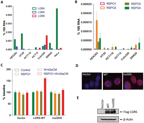
Background: LGR6 (leucine-rich repeat containing, G protein-coupled receptor 6) is a member of the rhodopsin-like seven transmembrane domain receptor superfamily with the highest homology to LGR4 and LGR5. LGR6 was found as one of the novel genes mutated in colon cancer through total exon sequencing and its promoter region is hypermethylated in 20-50% of colon cancer cases. In the skin, LGR6 marks a population of stem cells that can give rise to all cell lineages. Recently, we and others demonstrated that LGR4 and LGR5 function as receptors of R-spondins to potentiate Wnt/β-catenin signaling. However, the binding affinity and functional response of LGR6 to R-spondins, and the activity of colon cancer mutants of LGR6 have not been determined.

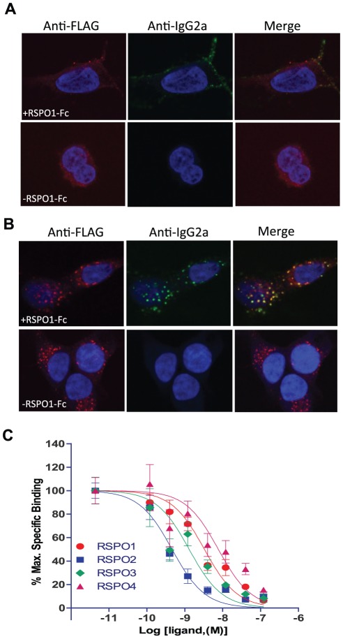
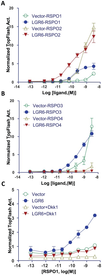
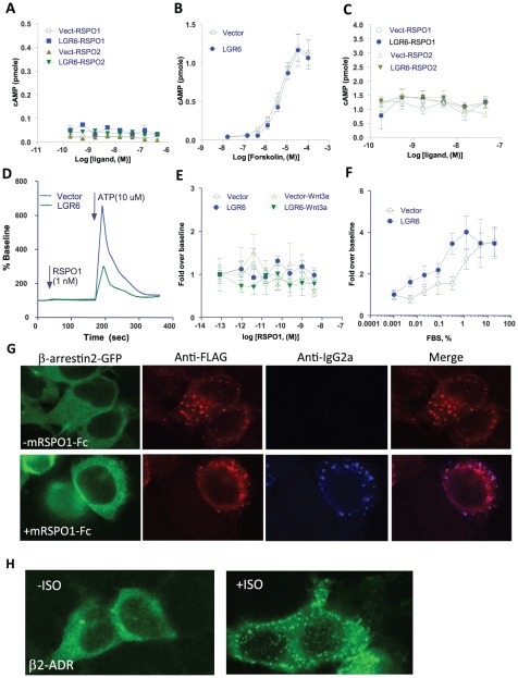
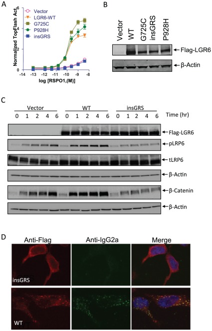
Principal findings: We found that LGR6 also binds and responds to R-spondins 1-3 with high affinity to enhance Wnt/β-catenin signaling through increased LRP6 phosphorylation. Similar to LGR4 and LGR5, LGR6 is not coupled to heterotrimeric G proteins or to β-arrestin following R-spondin stimulation. Functional and expression analysis of three somatic mutations identified in colon cancer samples indicates that one mutant fails to bind and respond to R-spondin (loss-of-function), but the other two have no significant effect on receptor function. Overexpression of wild-type LGR6 in HeLa cells leads to increased cell migration following co-treatment with R-spondin1 and Wnt3a when compared to vector control cells or cells overexpressing the loss-of-function mutant.

Conclusions: LGR6 is a high affinity receptor for R-spondins 1-3 and potentially functions as a tumor suppressor despite its positive effect on Wnt/β-catenin signaling.


Conflict of interest statement

Competing Interests: The authors have declared that no competing interests exist.

Figures

Figure 1
Figure 1. Binding of R-spondins to LGR6 by confocal immunofluorescence analysis and competition binding assay.
A&B, HEK293 cells stably expressing Flag-LGR6 were incubated with mRSPO1-Fc or control conditioned media (CM) at 4°C (A), or at 37°C (B). Flag-LGR6 was detected with a Cy3-labeled anti-Flag antibody (red) and mRSPO1-Fc was detected using Alexa488-labeled anti-IgG2a antibody (green). Nuclei were counterstained with To-Pro-3 (blue). The data shown are from one of three independent experiments with similar results. C, Quantitative binding analysis using a whole-cell-based assay. HEK293 cells stably expressing Flag-LGR6 were incubated with mRSPO1-Fc plus serial dilutions of purified recombinant RSPO1–4. Maximum specific binding is defined by the difference between the data with and without mRSPO1-Fc, which is ∼50% of total binding in general. All error bars are SEM (n = 3).
Figure 2
Figure 2. LGR6 enhances Wnt/β-catenin signaling in response to RSPO1–3 and this activity is inhibited by Dkk1.
HEK293T cells were transiently transfected with LGR6 or vector control, plus Dkk1 expression plasmids as indicated, along with the β-catenin reporter plasmid super 8× TOPFlash (firefly luciferase) and pRL-SV40 (renilla luciferase) and then stimulated with serial dilutions of purified recombinant RSPO1–2 (A), RSPO3–4 (B) or RSPO1 (C) in the presence of Wnt3a CM. Firefly luciferase activity of each well was normalized to that of renilla luciferase activity of the same well. All error bars are SEM (n = 4).
Figure 3
Figure 3. LGR6 is not coupled to heterotrimeric G proteins or to β-arrestin following R-spondin stimulation.
A, No cAMP response was detected in vector and LGR6 cells treated with RSPO1–2. B, Forksolin showed a strong stimulation of cAMP production in both cells. C, RSPO1–2 treatments had no effect on forskolin-stimulated cAMP production in vector and LGR6 cells. D, No Ca2+ mobilization was induced in vector and LGR6 cells in response to RSPO1, whereas ATP gave a robust response in both cells. E, Stimulation of LGR6 and vector cells by RSPO1 had no effect in the Gα(12/13) pathway using the serum response factor reporter enzyme assay. F, FBS gave a dose-dependent response in the serum response factor reporter enzyme assay. G, No translocation of β-arrestin was observed in LGR6 cells treated with mRSPO1-Fc, whereas colocalization of mRSPO1-Fc with LGR6 was confirmed. H, Robust translocation of β-arrestin was observed in HEK293 cells transfected with β2-adrenergic receptors and stimulated with its agonist isoproterenol.
Figure 4
Figure 4. Functional and binding analyses of LGR6 mutants.
A, TOPFlash assay of LGR6 mutants on RSPO1-induced potentiation of Wnt/β-catenin signaling. HEK293T cells were transiently transfected with expression plasmids as indicated with a combination of super 8× TOPFlash and pRL-SV40 reporter gene constructs, and then stimulated with serial dilutions of purified recombinant RSPO1 in the presence of Wnt3a CM. All error bars are SEM (n = 4). B, Expression levels of LGR6 mutants and WT in HEK 293T cells in transient transfection paradigms. Total cell lysates were treated with Laemmli buffer for 1 hr at 37°C, fractionated by SDS-PAGE using 4–20% gels, electrophoretically transferred to nitrocellulose membrane, and then probed with anti-Flag antibody. The signal was detected by ECL Western blotting detection reagents (Amersham Biosciences). β-actin was also probed as loading control. C, Time course of changes in Wnt3a-RSPO1-induced β-catenin accumulation and LRP6 phosphorylation in vector, LGR6-WT and insGRS-overexpressing cells. HEK293 cells stably expressing vector, LGR6-WT or insGRS were stimulated with 1 ng/ml RSPO1 plus Wnt3a CM for 0–6 hrs. Total cell lysates were probed with antibodies against Flag-LGR6, phosphor-Ser1490, total LRP6, and β-actin. For the analysis of nonmembrane-bound β-catenin, the cell lysates were cleared with ConA-sepharose beads and then probed with an antibody against β-catenin as described before . D, mRSPO1-Fc binding to LGR6-insGRS and WT. HEK293 cells stably expressing Flag-LGR6-WT or insGRS were incubated with mRSPO1-Fc at 37°C for 1 hr. The cells were fixed, permeabilized, and then co-stained with anti-Flag (red) and anti-IgG2a (green) antibodies to detect LGR6 and mRSPO1-Fc, respectively. Nuclei (blue) were counterstained with To-Pro-3.
Figure 5
Figure 5. Expression profiling of LGR4–6 and RSPO1–4 and migration assay of HeLa cells overexpressing LGR6.
A, Expression levels of LGR4–6 in HEK293 cells and a panel of cancer cell lines determined by quantitative RT-PCR analysis. B, Expression levels of RPSO1–4 in HEK293 and a panel of colon cancer cell lines determined by qPCR analysis. C, Cell migration analysis of HeLa cells stably expressing vector, LGR6-WT or insGRS after treatments with vehicle control, RSPO1,Wnt3a-CM, or RSPO1+Wnt3a. Data are presented at mean ± S.E.M. of three replicates after normalization to vehicle control. The experiment was repeated once and both showed similar results. D & E, Expression analysis of LGR6-WT and insGRS in HeLa cells by confocal immunofluorescence (D) and immunblotting (E) using an anti-Flag antibody.

References

    1. Hsu SY, Kudo M, Chen T, Nakabayashi K, Bhalla A, et al. The three subfamilies of leucine-rich repeat-containing G protein-coupled receptors (LGR): identification of LGR6 and LGR7 and the signaling mechanism for LGR7. Mol Endocrinol. 2000;14:1257–1271. - PubMed
    1. Barker N, Clevers H. Leucine-rich repeat-containing G-protein-coupled receptors as markers of adult stem cells. Gastroenterology. 2010;138:1681–1696. - PubMed
    1. Snippert HJ, Haegebarth A, Kasper M, Jaks V, van Es JH, et al. Lgr6 marks stem cells in the hair follicle that generate all cell lineages of the skin. Science. 2010;327:1385–1389. - PubMed
    1. Jaks V, Barker N, Kasper M, van Es JH, Snippert HJ, et al. Lgr5 marks cycling, yet long-lived, hair follicle stem cells. Nat Genet. 2008;40:1291–1299. - PubMed
    1. Barker N, van Es JH, Kuipers J, Kujala P, van den Born M, et al. Identification of stem cells in small intestine and colon by marker gene Lgr5. Nature. 2007;449:1003–1007. - PubMed

Publication types

MeSH terms