. 2012;7(5):e37281.
doi: 10.1371/journal.pone.0037281. Epub 2012 May 16.

A rat model of central venous catheter to study establishment of long-term bacterial biofilm and related acute and chronic infections

Affiliations

A rat model of central venous catheter to study establishment of long-term bacterial biofilm and related acute and chronic infections

Ashwini Chauhan et al. PLoS One. 2012.

Abstract

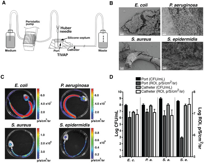
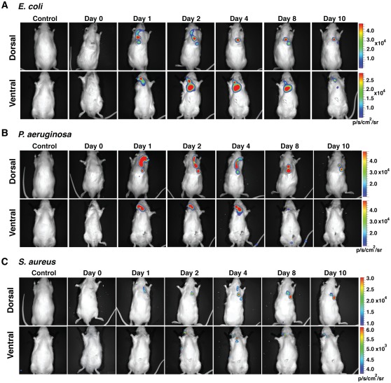
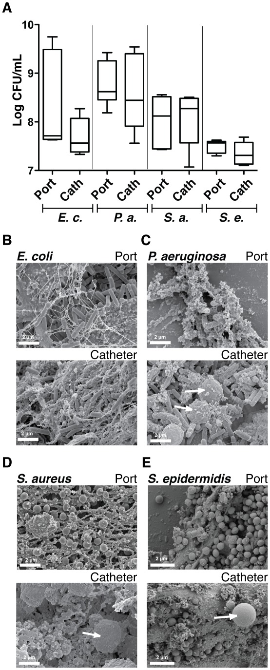
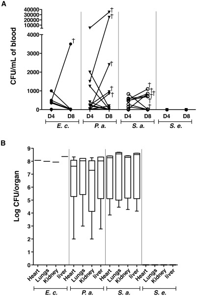
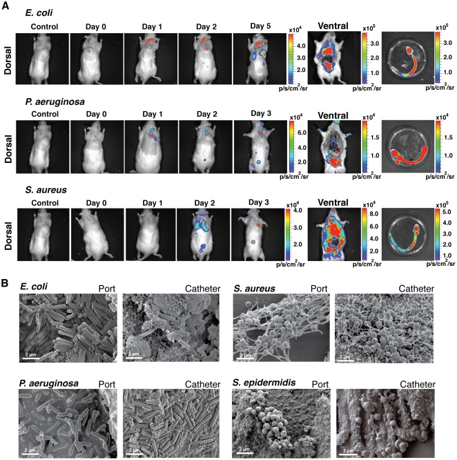
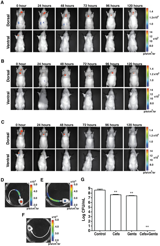
Formation of resilient biofilms on medical devices colonized by pathogenic microorganisms is a major cause of health-care associated infection. While in vitro biofilm analyses led to promising anti-biofilm approaches, little is known about their translation to in vivo situations and on host contribution to the in vivo dynamics of infections on medical devices. Here we have developed an in vivo model of long-term bacterial biofilm infections in a pediatric totally implantable venous access port (TIVAP) surgically placed in adult rats. Using non-invasive and quantitative bioluminescence, we studied TIVAP contamination by clinically relevant pathogens, Escherichia coli, Pseudomonas aeruginosa, Staphylococcus aureus and Staphylococcus epidermidis, and we demonstrated that TIVAP bacterial populations display typical biofilm phenotypes. In our study, we showed that immunocompetent rats were able to control the colonization and clear the bloodstream infection except for up to 30% that suffered systemic infection and death whereas none of the immunosuppressed rats survived the infection. Besides, we mimicked some clinically relevant TIVAP associated complications such as port-pocket infection and hematogenous route of colonization. Finally, by assessing an optimized antibiotic lock therapy, we established that our in vivo model enables to assess innovative therapeutic strategies against bacterial biofilm infections.


Conflict of interest statement

Competing Interests: The authors have declared that no competing interests exist.

Figures

Figure 1
Figure 1. Monitoring of in vitro biofilm-forming capacity of clinically relevant strains.
(A) in vitro continuous flow system to grow biofilms inside TIVAP. After 24 h biofilm formation in TIVAP was confirmed by scanning electron microscopy (SEM) for the four bacteria (B) and bioluminescence activity was acquired using an IVIS 100 camera (C). (D) Biofilm was analyzed by plating CFU/mL (n = 3 for each bacteria) on LB agar (E.c., E. coli or P.a., P. aeruginosa) or TSB agar (S.a., S. aureus or S.e., S. epidermidis) plates. Relative luminescence was correlated with CFU/mL by quantitative analysis of luminescence signals in the port and catheters separately using Living Image software.
Figure 2
Figure 2. Monitoring of TIVAP colonization by E. coli, P. aeruginosa and S. aureus.
The port was implanted subcutaneously at the dorsal midline and the catheter was inserted into the external jugular vein. Optimized doses of 104 CFU (E. coli, n = 5) or 106 CFU (P. aeruginosa, n = 7 and S. aureus, n = 6) in 100 µL were injected into the port and photon emission measured over a period of 10 days to monitor biofilm growth. Dorsal and ventral views of rats, showing progression of biofilm signals towards the catheter tip. Biofilm-associated infection was restricted to TIVAP by day 10. Control rat was catheterized but without bacterial inoculation. A representative animal is shown.
Figure 3
Figure 3. Bacterial colonization of TIVAP leads to biofilm formation.
Rats were sacrificed 10 days post infection, TIVAP were removed aseptically and cells harvested from the catheter and port separately and plated on LB agar (E.c., E. coli n = 5 or P.a., P. aeruginosa n = 7) or TSB agar (S.a., S. aureus n = 6 or S.e., S. epidermidis n = 5) plates for CFU/mL (A). (B–E) SEM images confirming true biofilm formation in TIVAP in vivo in the port and catheter. Representative images are presented. White arrows indicate eukaryotic immune cells.
Figure 4
Figure 4. Clinically relevant complications associated with TIVAP biofilms.
(A) Peripheral blood was harvested on day 4 (D4) and day 8 (D8) post-inoculation and plated on LB agar (E.c., E. coli n = 10 or P.a., P. aeruginosa n = 18) or TSB agar (S.a., S. aureus n = 14 or S.e., S. epidermidis n = 5) plates for CFU/mL. Bacteria were cleared by day 8 in most rats except for a few that suffered from systemic infection and died. Each dot represents one animal and cross (†) signifies the dead animal. (B) Organs were aseptically removed after sacrificing animals on day 10. Graph includes only rats that suffered from systemic infection and died. No bacteria were detected in the case of S. epidermidis (S. e., n = 5), whereas 10% rats suffered from systemic infection and died due to E. coli (E. c., n = 1/10) or ∼30% due to P. aeruginosa (P. a., n = 6/18) and S. aureus (S. a., n = 5/14) biofilms associated with TIVAP. Data are presented as box-and-whisker plots as previously described in material and methods.
Figure 5
Figure 5. Immunosuppression led to fatal biofilm infection.
(A) 102 CFU in 100 µL of the different bacteria were injected into the port of TIVAP implanted in cyclophosphamide-treated rats (100 mg/kg day -4 and 50 mg/kg day -1 of inoculation) and photon emission was monitored up to the death of the animals on days 3/5 to evaluate biofilm formation and associated infection. TIVAP were aseptically removed from dead animals and photon emission measured for both the animal and the extracted TIVAP. Control rat was a catheterized and cyclophosphamide-treated but without bacterial inoculation. (B) SEM images of TIVAP (port and catheter) infected with 4 clinical strains used in this study. Number of rats (n) used in the experiment, n = 4 for each strain.
Figure 6
Figure 6. In vivo efficacy of ALT.
ALT was instilled in the implanted colonized TIVAP (0 h) and associated with systemic vancomycin to treat S. aureus biofilm colonization. ALT was renewed every 24 h and its efficacy was monitored as photon emissions. Rats (n = 4, for each treatment) were sacrificed after 120 h of treatment and analyzed. (A) 5 mg/mL cefazolin ALT. (B) 1 mg/mL gentamicin ALT. (C) Combined cefazolin and gentamicin ALT (1∶1 v/v). (D–E) TIVAP were aseptically removed and photon emission due to remnant biofilm measured. (D) Cefazolin-treated TIVAP, (E) gentamicin-treated TIVAP and (F) TIVAP treated with cefazolin and gentamicin combination. In (A) to (F) representative experiments are shown. (G) TIVAP was extracted after treatment and cells were harvested and plated for CFU/mL. All the values are mean +/− standard deviation. Statistical analysis was done using one-way analysis of variance (ANOVA) using Graphpad Prism version 5.0c. p value<0.05 considered significant, *** (p<0.0001), ** (p<0.001) and * (p<0.05).

References

    1. Hall-Stoodley L, Stoodley P. Evolving concepts in biofilm infections. Cellular Microbiology. 2009;11:1034–1043. - PubMed
    1. Lynch AS, Robertson GT. Bacterial and fungal biofilm infections. Annu Rev Med. 2008;59:415–428. - PubMed
    1. Høiby N, Bjarnsholt T, Givskov M, Molin S, Ciofu O. Antibiotic resistance of bacterial biofilms. Int J Antimicrob Agents. 2010;35:322–332. - PubMed
    1. Wagner C, Hänsch GM, Wentzensen A, Heppert V. Implant-associated post-traumatic osteomyelitis. Bacterial biofilms and the immune defence as protagonists of the local inflammatory process. Unfallchirurg. 2006;109:761–769. - PubMed
    1. Baddour LM, Epstein AE, Erickson CC, Knight BP, Levison ME, et al. Update on cardiovascular implantable electronic device infections and their management: a scientific statement from the American Heart Association. Circulation. 2010;121:458–477. - PubMed

Publication types

MeSH terms