. 2012;7(5):e37342.
doi: 10.1371/journal.pone.0037342. Epub 2012 May 17.

Highly sensitive in vitro methods for detection of residual undifferentiated cells in retinal pigment epithelial cells derived from human iPS cells

Affiliations

Highly sensitive in vitro methods for detection of residual undifferentiated cells in retinal pigment epithelial cells derived from human iPS cells

Takuya Kuroda et al. PLoS One. 2012.

Abstract

Human induced pluripotent stem cells (hiPSCs) possess the capabilities of self-renewal and differentiation into multiple cell types, and they are free of the ethical problems associated with human embryonic stem cells (hESCs). These characteristics make hiPSCs a promising choice for future regenerative medicine research. There are significant obstacles, however, preventing the clinical use of hiPSCs. One of the most obvious safety issues is the presence of residual undifferentiated cells that have tumorigenic potential. To locate residual undifferentiated cells, in vivo teratoma formation assays have been performed with immunodeficient animals, which is both costly and time-consuming. Here, we examined three in vitro assay methods to detect undifferentiated cells (designated an in vitro tumorigenicity assay): soft agar colony formation assay, flow cytometry assay and quantitative real-time polymerase chain reaction assay (qRT-PCR). Although the soft agar colony formation assay was unable to detect hiPSCs even in the presence of a ROCK inhibitor that permits survival of dissociated hiPSCs/hESCs, the flow cytometry assay using anti-TRA-1-60 antibody detected 0.1% undifferentiated hiPSCs that were spiked in primary retinal pigment epithelial (RPE) cells. Moreover, qRT-PCR with a specific probe and primers was found to detect a trace amount of Lin28 mRNA, which is equivalent to that present in a mixture of a single hiPSC and 5.0×10⁴ RPE cells. Our findings provide highly sensitive and quantitative in vitro assays essential for facilitating safety profiling of hiPSC-derived products for future regenerative medicine research.


Conflict of interest statement

Competing Interests: The authors have declared that no competing interests exist.

Figures

Figure 1
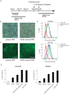
Figure 1. Differentiation of hiPSCs into retinal pigment epithelial cells.
(A) Schematic diagram of the culture procedure for retinal differentiation. Photomicrograph (B) and N-cadherin staining (C) shows that both primary RPE cells and hiPSC-derived RPE cells form polygonal, cobblestone-like morphology. Scale bars, 100 µm. (D) Flow cytometry analysis of CRALBP and GP-100 expression in hiPSCs (red), hiPSC-derived RPE cells (green) and primary RPE cells (blue). (E) Time-course analysis of expression of RPE cell markers, CRALBP (left) and RPE65 (right), using qRT-PCR. Error bars represent the standard deviation of the measurements (n = 3).
Figure 2
Figure 2. Soft agar colony formation assay of hiPSCs and teratocarcinoma PA-1 cells.
(A) Phase-contrast images of hiPSCs, primary RPE cells, hiPSC-derived RPE cells and PA-1cells spiked into primary RPE cells (1%) cultured in soft agar medium for 30 days. Arrows indicate the colonies of PA-1 cells. (B) PA-1 cells (1%, 100 cells; 0.5%, 50 cells; 0.25%, 25 cells; 0%, 0 cells) were spiked into 1.0×10⁴ primary RPE cells and grown in soft agar for 10, 20 and 30 days. Cell growth was quantified using a CytoSelect kit. Results were expressed as a relative fold change of the value of blank well. Statistical significance was determined using two-way ANOVA and Bonferroni's post-hoc test (*P<0.05 compared with the 0% control). (C) HiPSC-derived RPE cells, three lots of primary RPE cells and PA-1 cells spiked into primary RPE cells were grown in soft agar for 30 days. Quantification of the results is described in (B). Limit of detection was calculated as the mean plus 3.3 fold the standard deviation of the measurement of the three lots of primary RPE cells. Error bars represent the standard deviation of the measurements (n = 3).
Figure 3
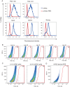
Figure 3. Detection of undifferentiated hiPSCs by flow cytometry assay.
(A) Flow cytometry analysis of hiPSCs (blue) and primary RPE cells (red). Cells were fixed, permiabilized and stained with anti-TRA-1-60, anti-TRA-1-81, anti-Sox2, anti-Oct3/4 and anti-Nanog antibodies labeled with fluorophore. (B) Five lots of primary RPE cells were analyzed by flow cytometry with anti-TRA-1-60 antibody. (C) HiPSCs (0.1%, 2.5×10² cells; 0.01%, 25 cells) were spiked into primary RPE cells (2.5×10⁵ cells) and analyzed by flow cytometry with anti-TRA-1-60 antibody. (D) Flow cytometry analysis of hiPSC-derived RPE cells was performed with anti-TRA-1-60 antibody. Ten thousand cells (A) and 1×10⁵ cells (B–D) were used for one assay of flow cytometry analysis. The numbers indicate the quantity of cells contained in the gate.
Figure 4
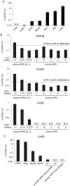
Figure 4. Detection of undifferentiated hiPSCs by qRT-PCR assay.
(A) The relative mRNA expressions in primary RPE cells of Lin28, Oct-3/4, Sox2, Nanog, Rex1, Klf4, and c-Myc were determined by qRT-PCR analysis. (B–D) qRT-PCR analysis of hiPSCs spiked into primary RPE cells and five lots of primary RPE cells. Single-cell hiPSCs (1%, 2.5×10³ cells; 0.1%, 2.5×10² cells; 0.01%, 25 cells) were spiked into 2.5×10⁵ primary RPE cells, and total RNA was isolated from the mixed cells. The mRNA levels of Nanog (B), Oct3/4 (C) and Lin28 (D) are shown as a relative expression. Limit of detection was calculated as the mean plus 3.3 fold the standard deviation of the measurement of the five lots of primary RPE cells. (E) Lin28 expression of hiPSCs differentiating into RPE and purified hiPSC-derived RPE cells (passage 3 and 4). All values are expressed as mRNA levels relative to those in undifferentiated hiPSCs. Results are means ± standard deviation (n = 3).

References

    1. Thomson JA, Itskovitz-Eldor J, Shapiro SS, Waknitz MA, Swiergiel JJ, et al. Embryonic stem cell lines derived from human blastocysts. Science. 1998;282:1145–1147. - PubMed
    1. Takahashi K, Tanabe K, Ohnuki M, Narita M, Ichisaka T, et al. Induction of pluripotent stem cells from adult human fibroblasts by defined factors. Cell. 2007;131:861–872. - PubMed
    1. Kehat I, Kenyagin-Karsenti D, Snir M, Segev H, Amit M, et al. Human embryonic stem cells can differentiate into myocytes with structural and functional properties of cardiomyocytes. J Clin Invest. 2001;108:407–414. - PMC - PubMed
    1. Zhang SC, Wernig M, Duncan ID, Brustle O, Thomson JA. In vitro differentiation of transplantable neural precursors from human embryonic stem cells. Nat Biotechnol. 2001;19:1129–1133. - PubMed
    1. Cai J, Zhao Y, Liu Y, Ye F, Song Z, et al. Directed differentiation of human embryonic stem cells into functional hepatic cells. Hepatology. 2007;45:1229–1239. - PubMed

Publication types

MeSH terms