. 2012 Jul;14(7):1772-87.
doi: 10.1111/j.1462-2920.2012.02778.x. Epub 2012 May 23.

Marine sulfate-reducing bacteria cause serious corrosion of iron under electroconductive biogenic mineral crust

Affiliations
Free PMC article

Marine sulfate-reducing bacteria cause serious corrosion of iron under electroconductive biogenic mineral crust

Dennis Enning et al. Environ Microbiol. 2012 Jul.
Free PMC article

Abstract

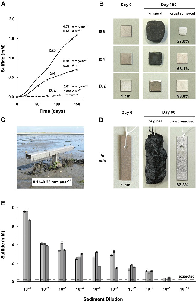
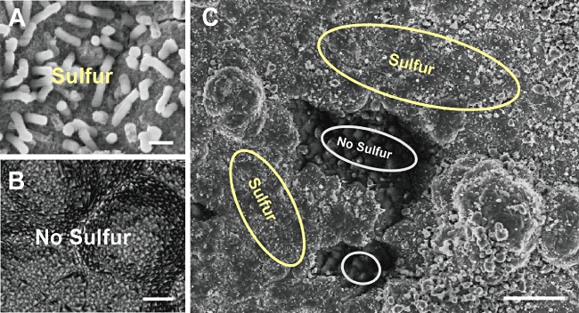
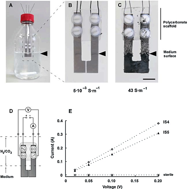
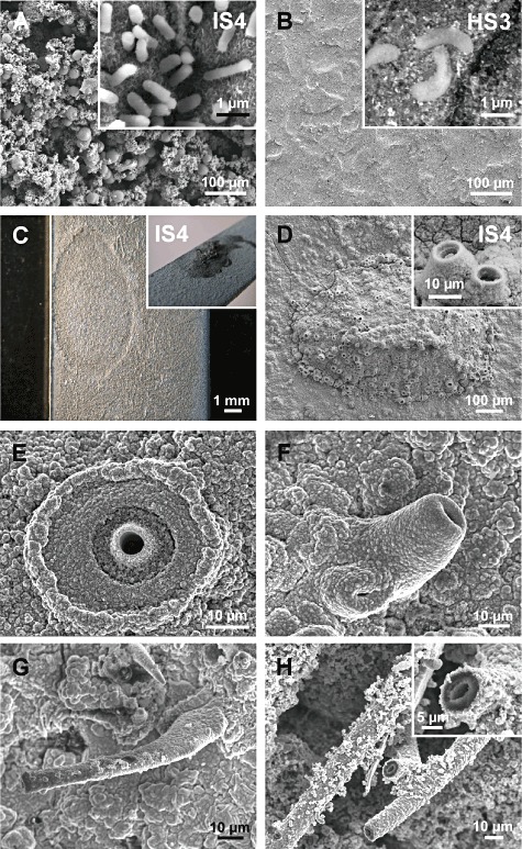
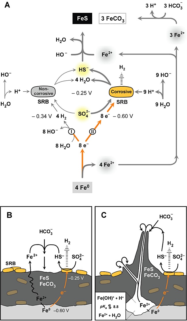
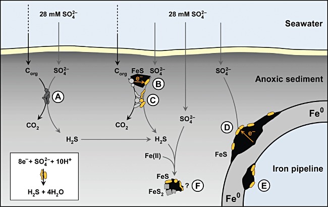
Iron (Fe(0) ) corrosion in anoxic environments (e.g. inside pipelines), a process entailing considerable economic costs, is largely influenced by microorganisms, in particular sulfate-reducing bacteria (SRB). The process is characterized by formation of black crusts and metal pitting. The mechanism is usually explained by the corrosiveness of formed H(2) S, and scavenge of 'cathodic' H(2) from chemical reaction of Fe(0) with H(2) O. Here we studied peculiar marine SRB that grew lithotrophically with metallic iron as the only electron donor. They degraded up to 72% of iron coupons (10 mm × 10 mm × 1 mm) within five months, which is a technologically highly relevant corrosion rate (0.7 mm Fe(0) year(-1) ), while conventional H(2) -scavenging control strains were not corrosive. The black, hard mineral crust (FeS, FeCO(3) , Mg/CaCO(3) ) deposited on the corroding metal exhibited electrical conductivity (50 S m(-1) ). This was sufficient to explain the corrosion rate by electron flow from the metal (4Fe(0) → 4Fe(2+) + 8e(-) ) through semiconductive sulfides to the crust-colonizing cells reducing sulfate (8e(-) + SO(4) (2-) + 9H(+) → HS(-) + 4H(2) O). Hence, anaerobic microbial iron corrosion obviously bypasses H(2) rather than depends on it. SRB with such corrosive potential were revealed at naturally high numbers at a coastal marine sediment site. Iron coupons buried there were corroded and covered by the characteristic mineral crust. It is speculated that anaerobic biocorrosion is due to the promiscuous use of an ecophysiologically relevant catabolic trait for uptake of external electrons from abiotic or biotic sources in sediments.


Figures

Fig. 1
Fig. 1
Corrosive sulfate-reducing bacteria in pure cultures and in situ. A. Long-term sulfide formation (measured as sulfate consumption) with iron coupons as the only electron donor in cultures of corrosive strains IS5 and IS4 (proposed: Desulfovibrio ferrophilus and Desulfopila corrodens respectively), and in a hydrogenotrophic control culture (Desulfopila inferna, D. i.). B. Thick corrosion crusts and metal loss in the same cultures. Residuary metal (% of initial) became obvious after crust removal by HCl-hexamine. C. Positioning device (stainless steel) for iron coupons in the Wadden Sea, island of Sylt (North Sea). Iron coupons were bound with threads to the device and buried for three months at ≥ 20 cm depth in anoxic sediment. D. Corrosion crust (with sand grains), and corroded metal (after crust dissolution) from (C). Here, the photographed fresh coupon (Day 0) is not the same as the incubated one. E. Sulfide formed (measured as sulfate consumed) after six months in serial dilutions (three in parallel) with native sediment (2 g, wet mass) from the same habitat. The line indicates sulfide expected solely by consumption of H2 formed from iron and seawater (based on independently measured H2-formation rates and experiments with merely H2-scavenging SRB).
Fig. 2
Fig. 2
X-ray microanalysis (EDX) of crust surface in a culture of corrosive strain IS4. A. Site colonized by cells (Bar, 1 µm). B. Site without microbial colonization (Bar, 1 µm). C. Both sites in the same field of view (Bar, 20 µm). Surface-attached cells of strain IS4 colocalize with the element S. Cells were not detectable at sulfur-free sites. Both, sulfur-containing and sulfur-free sites contained the elements Fe, C and O. Sulfur-free sites contained in addition Mg and Ca. Thirty point spectra at 10 kV were collected for each site. Resolution (lateral and vertical), 3–5 µm.
Fig. 3
Fig. 3
Determination of the conductivity of the corrosion crust. A. Anoxic bottle (600 ml medium) with two specially shaped fresh iron coupons. B. Coupons after three weeks of incubation in sterile medium. C. Coupons after three weeks of incubation with strain IS4. Bar, 1 cm. D. Scheme of the arrangement with voltage control and current measurement through separate circuits. E. Linear response of current to applied (non-electrolytic) voltage (0.02–0.2 V, DC).
Fig. 4
Fig. 4
Scanning electron micrographs of crust surface, colonization and microchimneys. A. Cells of strain IS4 on corrosion crust after three months. B. Cells of non-corrosive hydrogenotrophic control strain HS3 on slightly corroded metal surface after three months. C. Anodic iron dissolution (crust removed) underneath a ‘pustule’ (insert) formed by strain IS4 at high pH (≍ 9; Fig. 5C). D. ‘Pustule’ with short microchimneys (magnified in insert) in a culture of strain IS4. E. Microchimney developing from a crater-like structure in a culture of strain IS4. F. Short microchimney in a culture of strain IS4. G. Long microchimney in a culture of strain IS4. H. Long microchimney (magnified in insert) in an alkalizing corrosive enrichment.
Fig. 5
Fig. 5
Scheme of the stoichiometry and topology of direct (lithotrophic) corrosion. A. Stoichiometry of iron dissolution and channelling of electrons via H2 (I; classical scheme) or directly (II; new model) into sulfate reduction. Bold lines indicate the much faster electric ‘bypass’. Equilibrium redox potentials are indicated for real conditions at pH = 8 (see Appendix S1). Direct electron utilization provides a higher metabolic driving force (voltage: ΔE = 0.35 V, compared with 0.09 V via H2 of assumed ≍ 40 ppmv). Fe2+ not precipitated as sulfide may enter solution or precipitate with naturally widespread inorganic carbon as FeCO3. B. Electron flow through the crust to attached cells at pH = 8 (simplified, non-stoichiometric). Crust may contain co-precipitated calcium and magnesium carbonate, and/or cemented sand. The equivalent ion flow may occur via aqueous interstices (not depicted). C. Build-up of chimney-like ion bridges at pH ≥ 9. Reactions are essentially as in (B); however, there are pronounced spots of anodic iron dissolution. A significant pH gradient (low inside, high outside) causes Fe(II) precipitation at the rim, leading to chimney growth. Schemes include the possibility of H2 release (that may foster remote bacterial cells) due to an imbalance between electron uptake and sulfate reduction. Participation of possibly buried (encrusted) cells in sulfate reduction and H2 release is unknown.
Fig. 6
Fig. 6
Present synoptic hypotheses of the role of sulfate-reducing bacteria (SRB) capable of electron uptake from external sources in anoxic marine sediment (not to scale). A. Conventional organotrophic SRB. B. SRB interacting via electroconductive ferrous sulfide with electron-donating organotrophic anaerobic microorganisms. C. SRB interacting in direct contact with electron-donating organotrophic anaerobic microorganisms. D. Special SRB exploit metallic iron as electron source at the outer surface of a pipeline. E. Special SRB exploit metallic iron as electron source inside of a pipeline. F. Speculative possibility of pyritization of FeS (FeS + H2S → FeS2 + 2H+ + 2e) as a direct electron source for sulfate reduction.

References

    1. Appia-Ayme C, Guiliani N, Ratouchniak J, Bonnefoy V. Characterization of an operon encoding two c-type cytochromes, an aa3-type cytochrome oxidase, and rusticyanin in Thiobacillus ferrooxidans ATCC 33020. Appl Environ Microbiol. 1999;65:4781–4787. - PMC - PubMed
    1. Araujo JC, Teran FC, Oliveira RA, Nour EAA, Montenegro MAP, Campos JR, Vazoller RF. Comparison of hexamethyldisilazane and critical point drying treatments for SEM analysis of anaerobic biofilms and granular sludge. J Electron Microsc (Tokyo) 2003;52:429–433. - PubMed
    1. Auernik KS, Maezato Y, Blum PH, Kelly RM. The genome sequence of the metal-mobilizing, extremely thermoacidophilic archaeon Metallosphaera sedula provides insights into bioleaching-associated metabolism. Appl Environ Microbiol. 2008;74:682–692. - PMC - PubMed
    1. Badziong W, Thauer RK. Growth yields and growth rates of Desulfovibrio vulgaris (Marburg) growing on hydrogen plus sulfate and hydrogen plus thiosulfate as sole energy sources. Arch Microbiol. 1978;117:209–214. - PubMed
    1. Beech IB, Sunner IA. Sulphate-reducing bacteria and their role in corrosion of ferrous materials. In: Barton LL, Hamilton WA, editors. Sulphate-Reducing Bacteria: Environmental and Engineered Systems. Cambridge, UK: Cambridge University Press; 2007. pp. 459–482.

Publication types