. 2012 Sep 14;422(2):215-29.
doi: 10.1016/j.jmb.2012.05.021. Epub 2012 May 19.

Role of N-terminal myristylation in the structure and regulation of cAMP-dependent protein kinase

Affiliations

Role of N-terminal myristylation in the structure and regulation of cAMP-dependent protein kinase

Adam C Bastidas et al. J Mol Biol. .

Abstract

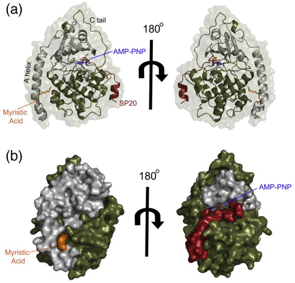
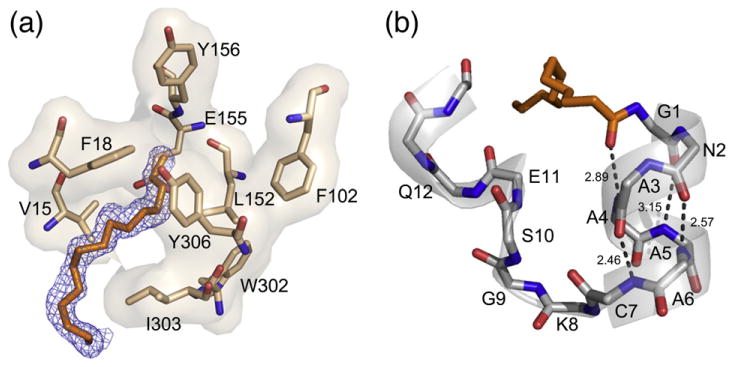
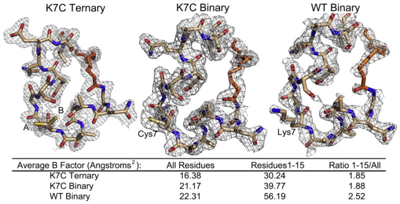
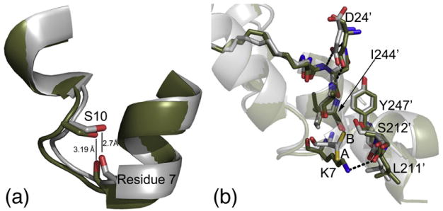
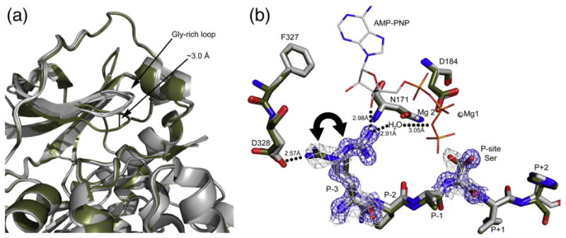
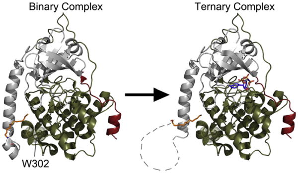
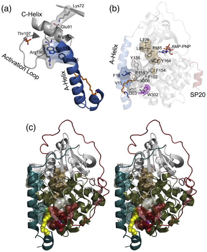
The catalytic (C) subunit of cAMP-dependent protein kinase [protein kinase A (PKA)] is a major target of cAMP signaling, and its regulation is of fundamental importance to biological processes. One mode of regulation is N-myristylation, which has eluded structural and functional characterization so far because most crystal structures are of the non-myristylated enzyme, are phosphorylated on Ser10, and generally lack electron density for the first 13 residues. We crystallized myristylated wild-type (WT) PKA and a K7C mutant as binary (bound to a substrate peptide) and ternary [bound to a substrate peptide and adenosine-5'-(β,γ-imido)triphosphate] complexes. There was clear electron density for the entire N-terminus in the binary complexes, both refined to 2.0 Å, and K7C ternary complex, refined to 1.35 Å. The N-termini in these three structures display a novel conformation with a previously unseen helix from residues 1 to 7. The K7C mutant appears to have a more stable N-terminus, and this correlated with a significant decrease in the B-factors for the N-terminus in the myr-K7C complexes compared to the WT binary complex. The N-terminus of the myristylated WT ternary complex, refined to 2.0 Å, was disordered as in previous structures. In addition to a more ordered N-terminus, the myristylated K7C mutant exhibited a 53% increase in k(cat). The effect of nucleotide binding on the structure of the N-terminus in the WT protein and the kinetic changes in the K7C protein suggest that myristylation or occupancy of the myristyl binding pocket may serve as a site for allosteric regulation in the C-subunit.


Conflict of interest statement

Conflict of Interest Statement. The authors report no conflict of interest.

Figures

Fig. 1
Fig. 1
The overall myristylated K7C ternary structure that displays the entire N-terminus in a novel conformation. (a) The overall structure of the myristylated K7C ternary complex is shown in cartoon representation with the small lobe colored gray, large lobe colored olive, SP20 colored red, AMP-PNP colored blue, and myristic acid colored orange. The protein surface is shown in transparent representation. This is the first time that the entire N-terminus and myristic acid are visible in a ternary complex and displays a novel α-helix from residues 1 to 7, unlike the WT ternary complex (see also Fig. S3). (b) The K7C ternary complex is shown as a surface representation with same color scheme as in (a). The surface representation highlights the myristic acid binding pocket as well as the platform created by the novel N-terminal helix.
Fig. 2
Fig. 2
The myristic acid binding pocket and N-terminal helix. (a) The location of the myristic acid and interacting residues within its binding pocket are shown in stick representation with the 2FoFc electron density map at 1 σ displayed for myristic acid in blue. (b) The myristic acid and main-chain atoms of the novel helix are shown in stick representation with H-bonding distances shown to highlight the properties of the helix.
Fig. 3
Fig. 3
Electron density of the N-termini for the structures displaying the entire N-terminus and myristic acid. The 2FoFc electron density maps contoured to 1 σ are shown for the first 15 residues and myristic acid for the myristylated K7C ternary, K7C binary, and WT binary structures. Residue 7 is annotated in each structure, and the K7C ternary structure adopts two conformations, A (65%) and B (35%). There is strong electron density for each structure at the N-terminus. However, the electron density for the WT binary structure is not as good with density lacking at some regions including part of the myristic acid and the Ser10 side chain. This is also reflected by the average B-factors for all residues compared to residues 1–15 for each structure that are listed in the figure.
Fig. 4
Fig. 4
Potential roles of the K7C mutation in the stabilization of the N-terminus. (a) The K7C (gray) and WT (olive) binary structures are aligned with the backbone atoms of residue 7 and side chain of Ser10 shown in stick representation. The distance between the Ser10 side chain and the backbone carbonyl of residue 7 is displayed, showing that the atoms are in H-bonding distance in the K7C mutant but not in the WT structure. It is possible that this hydrogen bond could stabilize the N-terminus. (b) K7C ternary and WT binary structures are aligned and colored as in (a); also shown here are the crystal packing interactions with symmetry-related molecules at the N-terminus. The K7C and WT structures adopt similar crystal packing with Ile210, Leu211, and Ser212, which are near the APE motif; Ile244 and Tyr247 from the G-helix; and Asp24 from the SP20 peptide. However, residue 7 and Leu211 from the symmetry-related molecule are slightly shifted in the WT structure possibly to facilitate packing and possibly to form a hydrogen bond between Lys7 and the backbone carbonyl of Leu211. Also, the K7C ternary structure adopts two conformations, A (65%) and B (35%), but the K7C binary structure only has density for conformation A. This B conformation would destabilize this crystal packing with the WT lysine residue.
Fig. 5
Fig. 5
Changes at the active site in the myristylated structures. (a) The four myristylated structures presented here are colored gray and aligned. Also aligned and displayed is another PKA structure, 1RDQ, in olive. The myristylated structures adopt a slightly raised glycine-rich loop, about 3 Å, compared to most closed PKA structures. (b) The active-site residues of the myr-K7C binary complex (olive) and myr-K7C ternary complex (gray) are shown. Also displayed is the location of the magnesium ions (gray spheres) and AMP-PNP (stick representation) from the ternary complex. The 2FoFc electron density maps at 1 σ are shown in gray for the binary complex and in blue for the ternary complex for the P-site Ser and P-3 Arg, highlighting the shifts in these residues upon formation of a ternary complex. The P-3 Arg was modeled facing the C-tail in the binary complexes, but there is some positive density for the P-3 Arg facing the active site, and it may exert some exchange between the two conformations. However, the residue only faces the active site in a ternary complex.
Fig. 6
Fig. 6
The formation of a ternary complex produces dynamic movements within the C-subunit that causes a disordering of the N-terminus. The C-subunit of PKA is shown in cartoon representation with the small lobe colored gray, large lobe colored olive, SP20 colored red, myristic acid colored orange, and AMP-PNP colored blue. The WT binary complex has a completely ordered N-terminus, which is displayed on the left, but upon formation of a ternary complex, the N-terminus of the WT C-subunit becomes disordered as illustrated on the right. The disordered regions of the N-terminus are displayed as connecting lines. Also, highlighted is Trp302, which is thought to be a sensor of active-site occupancy and may support the link between nucleotide binding and the conformation of the N-terminus.
Fig. 7
Fig. 7
Interactions of the myristic acid group and the A-helix within the C-subunit could influence the active site. (a) The A-helix is colored blue, myristic acid is colored orange, and other elements are colored gray. The A-helix residues Trp30 and Phe26 may interact with Arg93 and Arg190, which may influence the C-helix and the important Lys72–Glu91 interaction or the activation loop, respectively. (b) The A-helix is colored blue, myristic acid is colored orange, small lobe is colored gray, large lobe is colored olive, and SP20 is colored red. The protein is shown in transparent cartoon representation with specific residues highlighted in stick and surface representation. Residues in or near the myristate pocket may influence the active site such as Phe154 that is directly opposite of the myristate pocket and may influence the C-spine; residues Y164, F185, L95, and L106, which are colored brown; and the active site of the enzyme. Also, W302, highlighted in purple, is known to be a sensor of active-site occupancy and suggests potential cross-talk between the myristate pocket and active site. (c) A stereo view of the myristic acid pocket and interacting residues is displayed. In this depiction, the myristylated K7C binary structure is shown in cartoon representation with the N-terminus (residues 1–40) colored teal, the small lobe (residues 41–126) colored gray, the large lobe (residues 127–300) colored olive, the C-tail (residues 301–350) colored red, the myristic acid colored yellow and depicted in sphere representation, and the C-spine residues colored brown.

References

    1. Taylor SS, Kim C, Cheng CY, Brown SH, Wu J, Kannan N. Signaling through cAMP and cAMP-dependent protein kinase: diverse strategies for drug design. Biochim Biophys Acta. 2008;1784:16–26. - PMC - PubMed
    1. Kirschner LS, Yin Z, Jones GN, Mahoney E. Mouse models of altered protein kinase A signaling. Endocr-Relat Cancer. 2009;16:773–793. - PubMed
    1. Mucignat-Caretta C, Caretta A. Clustered distribution of cAMP-dependent protein kinase regulatory isoform RI alpha during the development of the rat brain. J Comp Neurol. 2002;451:324–333. - PubMed
    1. Francis SH, Corbin JD. Structure and function of cyclic nucleotide-dependent protein kinases. Annu Rev Physiol. 1994;56:237–272. - PubMed
    1. Tasken K, Skalhegg BS, Tasken KA, Solberg R, Knutsen HK, Levy FO, et al. Structure, function, and regulation of human cAMP-dependent protein kinases. Adv Second Messenger Phosphoprotein Res. 1997;31:191–204. - PubMed

Publication types

MeSH terms