

An anomalous segmental vein of the left upper lobe of the lung: preoperative identification by three-dimensional computed tomography pulmonary angiography
- PMID: 22617502
- PMCID: PMC3422930
- DOI: 10.1093/icvts/ivs205
An anomalous segmental vein of the left upper lobe of the lung: preoperative identification by three-dimensional computed tomography pulmonary angiography
Abstract
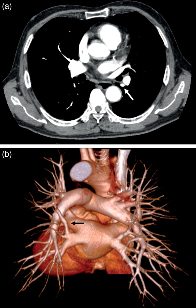
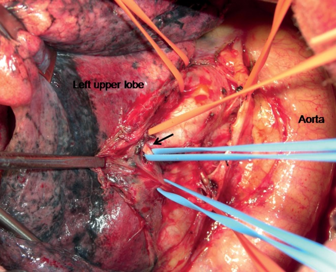
A number of variations in the pulmonary arteries and veins have been documented, and the information is very important for performing a safe lung resection. This report describes a case of an anomalous segmental vein of the left upper lobe of the lung. The patient was a 75-year old male who was suspected to have lung cancer in the left upper lobe. A contrast-enhanced computed tomography showed a vessel behind the left lower bronchus. A three-dimensional computed tomography angiography demonstrated that it was an anomalous vein for the apicoposterior segment of the left upper lobe of the lung, draining into the left inferior pulmonary vein. The aberrant vein was readily identified during surgery and was divided without injury, and a left upper lobectomy was successfully performed. Aberrant pulmonary veins for the superior segment of the right upper lobe of the lung are rarely observed, and the same kind of anomaly on the left side has not been reported.
Figures
References
-
- Kim J, Choi D, Lee K. CT of the bronchus intermedius: frequency and cause of a nodule in the posterior wall on normal scans. Am J Roentgenol. 1995;165:1349–52. - PubMed
-
- Asai K, Urabe N, Yajima K, Suzuki K, Kazui T. Right upper lobe venous drainage posterior to the bronchus intermedius: preoperative identification by computed tomography. Ann Thorac Surg. 2005;79:1866–71. - PubMed
-
- Watanabe S, Arai K, Watanabe T, Koda W, Urayama H. Use of three-dimensional computed tomographic angiography of pulmonary vessels for lung resections. Ann Thorac Surg. 2003;75:388–92. - PubMed
-
- Usami N, Iwao S, Mizuno T, Taniguchi T, Yokoi K. Three-dimensional angiography of aberrant segmental vein of right upper lobe. Asian Cardiovasc Thorac Ann. 2010;18:398. - PubMed
