Review
. 2012 Jul;24(4):408-16.
doi: 10.1097/BOR.0b013e32835461d3.

Streptococcus and rheumatic fever

Affiliations
Review

Streptococcus and rheumatic fever

Madeleine W Cunningham. Curr Opin Rheumatol. 2012 Jul.

Abstract

Purpose of review: To give an overview of the current hypotheses of the pathogenesis of rheumatic fever and group A streptococcal autoimmune sequelae of the heart valve and brain.

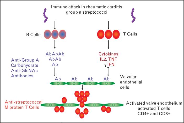
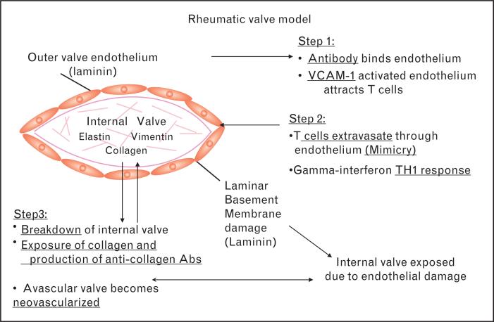
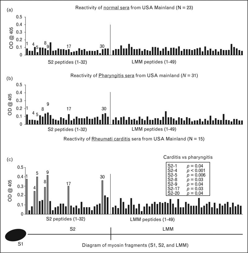
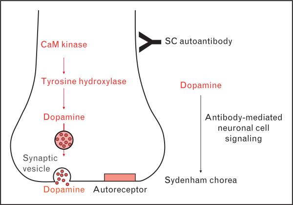
Recent findings: Human monoclonal antibodies (mAbs) derived from rheumatic heart disease have provided evidence for crossreactive autoantibodies that target the dominant group A streptococcal epitope of the group A carbohydrate, N-acetyl-beta-D-glucosamine (GlcNAc), and heart valve endothelium, laminin and laminar basement membrane. T cells in peripheral blood and in rheumatic heart valves revealed the presence of T cells crossreactive with streptococcal M protein and cardiac myosin. For initiation of disease, evidence suggests a two-hit hypothesis for antibody attack on the valve endothelium with subsequent extravasation of T cells through activated endothelium into the valve to form granulomatous lesions and Aschoff bodies. Autoantibodies against the group A streptococcal carbohydrate epitope GlcNAc and cardiac myosin and its peptides appear during progression of rheumatic heart disease. However, autoantibodies against collagen that are not crossreactive may form because of the release of collagen from damaged valve or to responses to collagen bound in vitro by certain serotypes of streptococci. In Sydenham chorea, human mAbs derived from disease target the group A carbohydrate epitope GlcNAc and gangliosides and dopamine receptors found on the surface of neuronal cells in the brain. Human mAbs and autoantibodies in Sydenham chorea were found to signal neuronal cells and activate calcium calmodulin-dependent protein kinase II (CaMKII) in neuronal cells and recognize the intracellular protein biomarker tubulin.

Summary: To summarize, pathogenic mechanisms of crossreactive autoantibodies which target the valve in rheumatic heart disease and the neuronal cell in Sydenham chorea share a common streptococcal epitope GlcNAc and target intracellular biomarkers of disease including cardiac myosin in the myocardium and tubulin, a protein abundant in the brain. However, intracellular antigens are not believed to be the basis for disease. The theme of molecular mimicry in streptococcal autoimmune sequelae is the recognition of targeted intracellular biomarker antigens such as cardiac myosin and brain tubulin, while targeting extracellular membrane antigens such as laminin on the valve surface endothelium or lysoganglioside and dopamine receptors in the brain. Antibody binding to these cell surface antigens may lead to valve damage in rheumatic heart disease or neuropsychiatric behaviors and involuntary movements in Sydenham chorea.


Figures

FIGURE 1
FIGURE 1
Two-hit hypothesis of initiation of rheumatic carditis. Group A streptococcal infection leads to the production of antigroup A carbohydrate antibody (B cells) which crossreacts with the valve endothelium and upregulates vascular cell adhesion molecule-1 (VCAM-1) on the valve endothelium in Step 1. In Step 2, T cells responsive to streptococcal M protein epitopes adhere to the VCAM-1 on activated valve surface endothelium and extravasate into the valve. The diagram illustrates the first two initial steps of rheumatic heart disease. Source: Similar but slightly different from figure in [34].
FIGURE 2
FIGURE 2
Multistep hypothesis of development of rheumatic carditis and heart disease. Diagram illustrating the process of initial mimicry which leads to granuloma formation, gamma interferon production and scarring in the valve. After the initial process of inflammation has developed in the valve, other proteins in the valve may then be recognized by the immune system leading potentially to epitope spreading and responses against other valve proteins such as vimentin and collagen. Source: Similar but wording on figure different from figure in [35]. Similar but slightly different from figure in [34].
FIGURE 3
FIGURE 3
Reactivity of serum IgG from rheumatic heart disease with human cardiac myosin peptides from the S2 and LMM rod regions in the enzyme-linked immunosorbent assay. (a) Mean reactivity of normal serum IgG from control donors with no evidence of streptococcal infection or heart disease on the U.S. mainland against S2 and LMM peptides. (b) Mean reactivity of serum IgG from patients with streptococcal pharyngitis on the US mainland against S2 and LMM peptides. (c) Serum IgG from patients with rheumatic carditis reacted with peptides S2–1, S2–4, S2–5, S2–8, S2–9, S2–17, and S2–30, compared with the reactivity of serum IgG from patients with pharyngitis against those same peptides (b). Unadjusted Mann–Whitney P values for the comparison between carditis and pharyngitis from the U.S. mainland are shown in panel (c). The comparison for S2–4 is statistically significant on the basis of a two-sided alpha level adjusted to preserve the false-discovery rate at 5% (1 : 100 dilution of serum). Data from [24■■].
FIGURE 4
FIGURE 4
Sydenham chorea-derived human monoclonal antibody (mAb) stimulated an increase in tyrosine hydroxylase synthesis in rat brain neurons. Sydenham chorea-derived mAb 24.3.1 was passively transferred intrathecally into rat brain and the increase in tyrosine hydroxylase was determined by immunohistochemistry. Chorea-derived mAb (24.3.1) induced higher levels of tyrosine hydroxylase (left figures – pink) in neurons compared with isotype control (right figures – blue). Insets show negative (blue) regions of the rat brain. The ability of chorea antibodies to alter neurotransmitter synthesis and release may explain the efficacy of dopamine receptor blockers such as haloperidol in the treatment of Sydenham's chorea. Data from [28].
FIGURE 5
FIGURE 5
Simplified illustration of a potential pathogenic mechanism in Sydenham chorea. Antineuronal antibody (IgG) may bind to receptors on neuronal cells and trigger the signaling cascade of CaMKII, tyrosine hydroxylase and dopamine release which may potentially lead to excess dopamine and the manifestations of Sydenham chorea. Source: Similar but slightly different from figure in [34].

Similar articles

Cited by

References

    1. Cunningham MW. Pathogenesis of group A streptococcal infections. Clin Microbiol Rev. 2000;13:470–511. - PMC - PubMed
    1. Stollerman GH, editor. Rheumatic fever and streptococcal infection. Grune and Stratton; New York: 1975. pp. 1–145.
    1. Taranta A, Stollerman GH. The relationship of Sydenham's chorea to infection with group A streptococci. Am J Med. 1956:170–175. - PubMed
    1. Stollerman GH. Rheumatic fever. Lancet. 1997;349:935–942. - PubMed
    1. Dajani AS. Guidelines for the diagnosis of rheumatic fever (Jones criteria 1992 update). J Am Med Assoc 1992. 268:2069–2073. - PubMed

Publication types