. 2012 Jun 5;109(23):9191-6.
doi: 10.1073/pnas.1120067109. Epub 2012 May 22.

Computational model of a circulation current that controls electrochemical properties in the mammalian cochlea

Affiliations

Computational model of a circulation current that controls electrochemical properties in the mammalian cochlea

Fumiaki Nin et al. Proc Natl Acad Sci U S A. .

Abstract

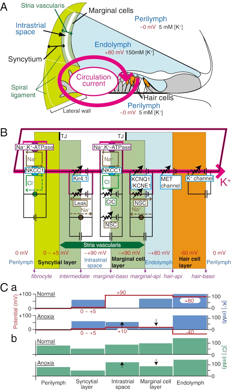
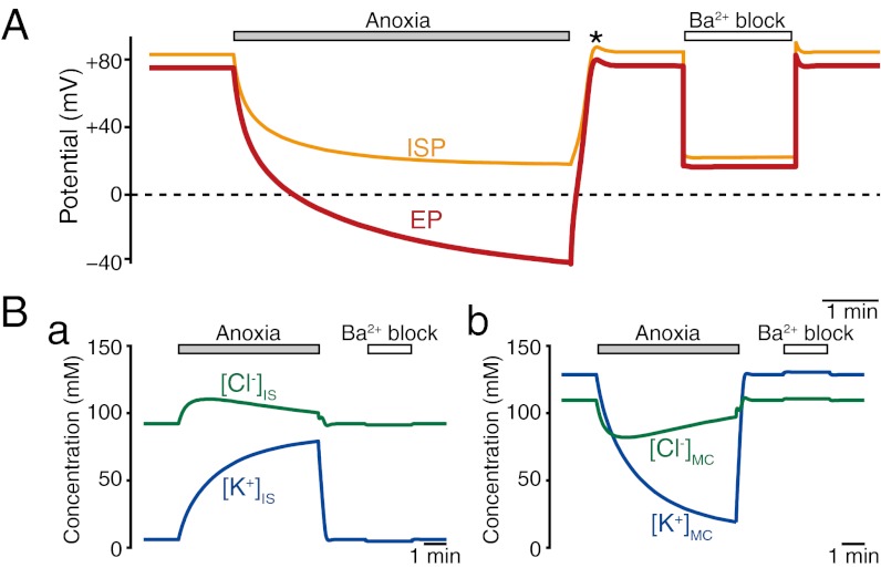
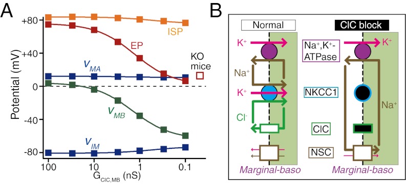
Sound-evoked mechanical stimuli permit endolymphatic K(+) to enter sensory hair cells. This transduction is sensitized by an endocochlear potential (EP) of +80 mV in endolymph. After depolarizing the cells, K(+) leaves hair cells in perilymph, and it is then circulated back to endolymph across the lateral cochlear wall. In theory, this process entails a continuous and unidirectional current carried by apical K(+) channels and basolateral K(+) uptake transporters in both the marginal cell and syncytial layers of the lateral wall. The transporters regulate intracellular and extracellular [K(+)], allowing the channels to form K(+) diffusion potentials across each of the two layers. These diffusion potentials govern the EP. What remains uncertain is whether these transport mechanisms accumulating across diverse cell layers make up a continuous circulation current in the lateral wall and how this current might affect the characteristics of the endolymph. To address this question, we developed an electrophysiological model that incorporates channels and transporters of the lateral wall and channels of hair cells that derive a circulation current. The simulation replicated normal experimental EP values and reproduced experimentally measured changes in the EP and intra- and extracellular [K(+)] in the lateral wall when different transporters and channels were blocked. The model predicts that, under these different conditions, the circulation current's contribution to the EP arises from different sources. Finally, our model also accurately simulated EP loss in a mouse model of a chloride channelopathy associated with deafness.


Conflict of interest statement

The authors declare no conflict of interest.

Figures

Fig. 1.
Fig. 1.
Key elements of the NHK model. (A) Structure of the cochlea and the concept underlying the circulation current. The circulation current (pink arrow) unidirectionally flows through a pathway comprising the endolymph, hair cells, perilymph, and lateral cochlear wall, and it flows back to the endolymph. In normal conditions, this flow would be carried by K+. (B) The electrical circuit and its components used in the modeling. The channels and transporters that are involved in driving the circulation current as well as the potential in each compartment are described. The fibrocytes belong to the spiral ligament (A). The stria vascularis contains three cell types: basal cells, intermediate cells, and marginal cells. Basal cells, which are omitted in this scheme, are apposed to the fibrocytes and intermediate cells. Note that the apical and basolateral surfaces of the syncytial layer correspond to the membranes of intermediate cells and fibrocytes, respectively. api, apical; baso, basolateral; ClC, ClC-type Cl channels; Leak, leak channels; MET, mechanoelectrical transduction; NKCC1, Na+, K+, 2 Cl cotransporter type 1; NSC, nonselective cation channels; TJ, tight junction. The circles filled with green and brown indicate the Cl and Na+ transporters, respectively. (C) Summary profile of [K+] (a; blue bars), [Cl] (b; green bars), and potential (a; red lines and numbers) of the lateral wall on the basis of the experiments in the work by Nin et al. (18) and Figs. S2A and S6C. The EP primarily represents the sum of two K+ diffusion potentials across the apical membranes of the syncytial and marginal cell layers (C, a Upper) (18). C, a Lower shows that anoxia causes [K+] in the IS to increase and [K+] in marginal cells to decrease, modulating the two K+ diffusion potentials. Consequently, the EP falls to a negative value. Anoxia also changes [Cl] properties (C, b). In any condition, the potential of the syncytial layer is constant. In C, a and b, up and down arrows during anoxia (Lower) indicate increase and decrease of [K+] or [Cl], respectively, compared with normal conditions (Upper).
Fig. 2.
Fig. 2.
Simulation of electrochemical properties of the lateral wall and endolymph. Traces exhibit the calculated results of the ISP (orange line in A), EP (red line in A), [K+] in IS and marginal cells ([K+]IS and [K+]MC; blue lines in B, a and b), and [Cl] in the IS and marginal cells ([Cl]IS and [Cl]MC; green lines in B, a and b) in normal, anoxic, and Ba2+ block conditions. In this figure and subsequent figures (Fig. 3A), the initial values of all of the potentials and concentrations were steady-state values that developed over the course of 3.5 min after the start of the simulation (for the values in the initial 4 min, see Fig. S5 C and D). The asterisk in A marks the overshoots of the potentials after a period of anoxia (18).
Fig. 3.
Fig. 3.
Simulated membrane potentials and currents in the lateral wall. (A) Responses of the membrane potentials in anoxic and Ba2+ block conditions (solid lines) (Fig. S5C). vMA, vMB, and vIM indicate the potentials across the apical and basolateral membranes of marginal cells and intermediate cells' membranes, respectively. Note that intermediate cells provide the apical surface of the syncytial layer. The purple dotted lines illustrate the change of K+ equilibrium potentials (EK) across the apical membranes of marginal cells (Upper) and intermediate cells' membranes (Lower), which are calculated from [K+] in endolymph (Table S2) and marginal cells (Fig. 2B, b). In the IS (Fig. 2B, a) and syncytial layer (Table S2), they are calculated by the relation EK = RT/F([K+]o/[K+]i), where [K+]i and [K+]o are intra- and extracellular [K+] adjacent to the membrane, respectively. (B) The behavior of the circulation current (pink). (C) The net K+, Na+, and Cl currents across the basolateral membranes of marginal cells. Each current is a sum of the respective ionic flux carried by the channels and transporters on the membranes (Fig. S7). The circulation current (pink) underlies the K+ current (blue) for comparison. The currents shown in this figure are derived from analysis of the data during the periods marked with the top bars in A and B.
Fig. 4.
Fig. 4.
Effects of dysfunction of Cl flow of the lateral wall. Plotted are the steady-state values of the EP, ISP, and membrane potentials (Fig. 3A describes abbreviations) as a function of the conductance of ClC-type Cl channels (A). These potentials represent values that were predicted for a time point 18,000 s into the simulation. This ensured that all potentials represented steady-state values, therefore mimicking unperturbed values that would be measured in living animals that have varying degrees of ClC conductance. The EP measured in barttin KO mice, which lack functional ClC channels (10), is indicated by the open square. B illustrates the predicted ionic flows across the basolateral membranes of marginal cells in the normal condition (Left) and during block of ClC channels (Right). Under normal conditions, NKCC1 cancels the Cl flow through ClC channels. During block of ClC channels, Na+ uptake through the NKCC1 is reduced; consequently, Na+ inflow is rerouted from NKCC1 to NSC channels, hyperpolarizing vMB (details in SI Text). The symbols filled with black indicate the dysfunction of the channels or transporters that is predicted to occur.

References

    1. Bekesy G. Resting potentials inside the cochlear partition of guinea pig. Nature. 1952;169:241–242. - PubMed
    1. Hudspeth AJ. How the ear’s works work. Nature. 1989;341:397–404. - PubMed
    1. Davis H. Some principles of sensory receptor action. Physiol Rev. 1961;41:391–416. - PubMed
    1. Wangemann P, Schacht J. Homeostasic mechanisms in the cochlea. In: Dallos P, Popper AN, Fay R, editors. Springer Handbook of Auditory Research: The Cochlea. New York: Springer; 1996.
    1. Wangemann P. Supporting sensory transduction: Cochlear fluid homeostasis and the endocochlear potential. J Physiol. 2006;576:11–21. - PMC - PubMed

Publication types

LinkOut - more resources