. 2012 Jun;139(12):2150-5.
doi: 10.1242/dev.077065.

Cellular retinoic acid-binding proteins are essential for hindbrain patterning and signal robustness in zebrafish

Affiliations

Cellular retinoic acid-binding proteins are essential for hindbrain patterning and signal robustness in zebrafish

Anna Q Cai et al. Development. 2012 Jun.

Abstract

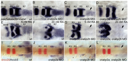
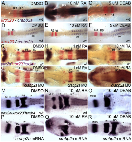
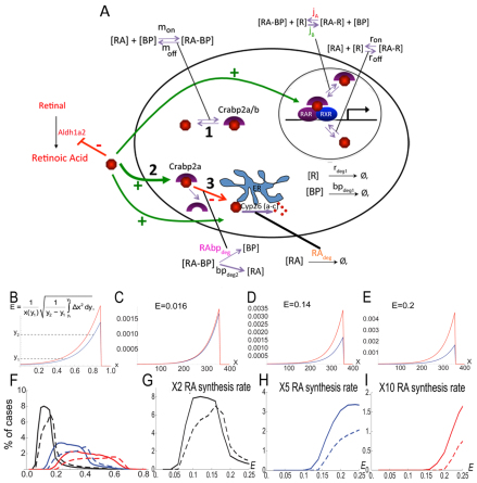
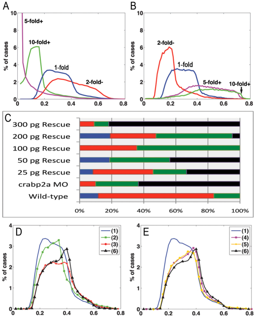
The vitamin A derivative retinoic acid (RA) is a morphogen that patterns the anterior-posterior axis of the vertebrate hindbrain. Cellular retinoic acid-binding proteins (Crabps) transport RA within cells to both its nuclear receptors (RARs) and degrading enzymes (Cyp26s). However, mice lacking Crabps are viable, suggesting that Crabp functions are redundant with those of other fatty acid-binding proteins. Here we show that Crabps in zebrafish are essential for posterior patterning of the hindbrain and that they provide a key feedback mechanism that makes signaling robust as they are able to compensate for changes in RA production. Of the four zebrafish Crabps, Crabp2a is uniquely RA inducible and depletion or overexpression of Crabp2a makes embryos hypersensitive to exogenous RA. Computational models confirm that Crabp2a improves robustness within a narrow concentration range that optimizes a 'robustness index', integrating spatial information along the RA morphogen gradient. Exploration of signaling parameters in our models suggests that the ability of Crabp2a to transport RA to Cyp26 enzymes for degradation is a major factor in promoting robustness. These results demonstrate a previously unrecognized requirement for Crabps in RA signaling and hindbrain development, as well as a novel mechanism for stabilizing morphogen gradients despite genetic or environmental fluctuations in morphogen availability.


Figures

Fig. 1.
Fig. 1.
crabp2a and crabp2b are required for zebrafish hindbrain patterning. (A-L) Whole-mount in situ hybridization (ISH) for pax2a at the midbrain-hindbrain boundary (MHB), krox20 in rhombomere (R) 3 and R5, and hoxb4 (A-H) or hoxb5 (I-L) in R7 and spinal cord at 18 hpf; dorsal views, anterior left. Compared with uninjected controls (A), morpholino (MO) depletion of Crabp2a (B,J) or Crabp2b (C,K) slightly reduces and co-injection (D,L) eliminates hoxb4 and hoxb5 expression (arrows). Exogenous RA treatments (5-20 nM) that posteriorize the hindbrain do not rescue hoxb4 expression (E-H). wt, wild type.
Fig. 2.
Fig. 2.
crabp2a is RA inducible and required for signal robustness. (A-F) Whole-mount ISH for crabp2a/b expression at 15 hpf; dorsal views, anterior left. crabp2a (A-C) is expressed posterior to the R5/6 boundary, whereas crabp2b (D-F) expression is restricted to somites. Treatment with 10 nM RA expands crabp2a expression anteriorly throughout the CNS (B) but has no effect on crabp2b (E). Treatment with 5 μM DEAB eliminates crabp2a expression (C) but does not disrupt crabp2b (F). (G-R) pax2a, krox20 and hoxb4 expression at 18 hpf in uninjected controls (G-I), Crabp2a-MO-injected (J-L) and crabp2a mRNA-injected embryos (M-R) treated with 1 nM RA (H,K), 10 nM RA (I,L,N,Q) or 10 μM DEAB (O,R). so, somites.
Fig. 3.
Fig. 3.
Computational models indicate essential roles for Crabps in robustness. (A) Schematic of RA signaling in one cell illustrating model components: RA (red), Crabps (purple crescents), Cyp26s associated with endoplasmic reticulum (ER, blue), RA-degradation products ([deg], red dots). Green arrows indicate components induced by RA and red inhibitory symbols indicate components repressed by RA. (B) A robustness index (E) utilized: where ymin is the minimum value and yref(xref) is an imposed maximum value of the reference gradient. (C-E) Three examples of calculated gradients very similar (C) or different (D,E) from the reference gradient (red line) across the hindbrain primordium (x-axis) and corresponding E values. (F-I) Probability density distributions (percentages, y-axis) of E values (x-axis) for models that either include binding proteins (solid lines) or do not (dashed lines) for models with a 2-fold (black lines, G), 5-fold (blue lines, H) or 10-fold (red lines, I) increase in [RA]out synthesis rate.
Fig. 4.
Fig. 4.
Robustness gains require Crabp-mediated RA degradation. (A,B) E value distributions for models with either a 5-fold increase (A) or decrease (B) of RA synthesis rate with respect to 10- (green), 5- (magenta), or 2-fold (red) increases in Vbp. (C) Percentages (x-axis) of wild-type (blue bars), mild (red bars), moderate (green bars) and severe (black bars) phenotypes resulting from injection of different amounts of Crabp2a mRNA per embryo (y-axis) treated with 5 nM RA. (D,E) Simulations of 5-fold increases in RA synthesis rate in which binding proteins: (1) have all three mechanisms; (2) do not transport RA to receptors (jB=0); (3) do not sequester RA (jA=0); (4) do not transport RA to Cyp26 degradation (rabpdeg=0); (5) must bind RA to allow degradation (radeg=0); and (6) are absent.

References

    1. Allenby G., Janocha R., Kazmer S., Speck J., Grippo J. F., Levin A. A. (1994). Binding of 9-cis-retinoic acid and all-trans-retinoic acid to retinoic acid receptors alpha, beta, and gamma. Retinoic acid receptor gamma binds all-trans-retinoic acid preferentially over 9-cis-retinoic acid. J. Biol. Chem. 269, 16689–16695 - PubMed
    1. Aström A., Tavakkol A., Pettersson U., Cromie M., Elder J. T., Voorhees J. J. (1991). Molecular cloning of two human cellular retinoic acid-binding proteins (CRABP). Retinoic acid-induced expression of CRABP-II but not CRABP-I in adult human skin in vivo and in skin fibroblasts in vitro. J. Biol. Chem. 266, 17662–17666 - PubMed
    1. Aström A., Pettersson U., Chambon P., Voorhees J. J. (1994). Retinoic acid induction of human cellular retinoic acid-binding protein-II gene transcription is mediated by retinoic acid receptor-retinoid X receptor heterodimers bound to one far upstream retinoic acid-responsive element with 5-base pair spacing. J. Biol. Chem. 269, 22334–22339 - PubMed
    1. Begemann G., Schilling T. F., Rauch G. J., Geisler R., Ingham P. W. (2001). The zebrafish neckless mutation reveals a requirement for raldh2 in mesodermal signals that pattern the hindbrain. Development 128, 3081–3094 - PubMed
    1. Ben-Zvi D., Barkai N. (2010). Scaling of morphogen gradients by an expansion-repression integral feedback control. Proc. Natl. Acad. Sci. USA 107, 6924–6929 - PMC - PubMed

Publication types

MeSH terms