. 2012:2012:826178.
doi: 10.1155/2012/826178. Epub 2012 Feb 12.

Prevention of chronic experimental colitis induced by dextran sulphate sodium (DSS) in mice treated with FR91

Affiliations

Prevention of chronic experimental colitis induced by dextran sulphate sodium (DSS) in mice treated with FR91

Valter R M Lombardi et al. J Biomed Biotechnol. 2012.

Abstract

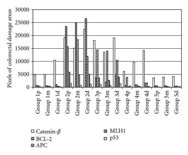
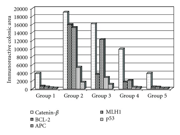
One of the main treatments currently used in humans to fight cancer is chemotherapy. A huge number of compounds with antitumor activity are present in nature, and many of their derivatives are produced by microorganisms. However, the search for new drugs still represents a main objective for cancer therapy, due to drug toxicity and resistance to multiple chemotherapeutic drugs. In animal models, a short-time oral administration of dextran sulfate sodium (DSS) induces colitis, which exhibits several clinical and histological features similar to ulcerative colitis (UC). However, the pathogenic factors responsible for DSS-induced colitis and the subsequent colon cancer also remain unclear. We investigated the effect of FR91, a standardized lysate of microbial cells belonging to the Bacillus genus which has been previously shown to have significant immunomodulatory effects, against intestinal inflammation. Colitis was induced in mice during 5 weeks by oral administration 2% (DSS). Morphological changes in the colonic mucosa were evaluated by hematoxylin-eosin staining and immunohistochemistry methods. Adenocarcinoma and cryptal cells of the dysplastic epithelium showed cathenin-β, MLH1, APC, and p53 expression, together with increased production of IFN-γ. In our model, the optimal dose response was the 20% FR91 concentration, where no histological alterations or mild DSS-induced lesions were observed. These results indicate that FR91 may act as a chemopreventive agent against inflammation in mice DSS-induced colitis.


Figures

Figure 1
Figure 1
Experimental protocol for DSS-induced colitis/colon cancer in mice and the administration of FR91 at different concentrations.
Figure 2
Figure 2
Histological sections of three different portions of the experimental mice colon to show histopathological lesions identified by hematoxylin and eosin staining. (a–c) Transverse sections at three different colorectal levels (proximal, middle, and distal) of mice group 1 treated with FR91 (10%). Note the absence of colonic lesions and the similar epithelial structures observed in normal control mice. (d–f) Transverse sections at three different colorectal levels of mice group 2 treated with DSS (2%) for 5 weeks. In each studied level, multiple histopathological lesions were observed, being particularly clear the severe-grade dysplasia (arrowheads in (d) and (e)) developed in the colon of mice from this group, together with aberrant crypts (arrow in (d)), adenomatous polyps (arrowhead in (f)), and incipient ulcerations (arrow in (f)). (g–I) Transverse sections at three different colorectal levels of mice group 3, with the administration of DSS (2%) for 5 weeks and treated with FR-91 (5%). Although these three colonic levels show a low-grade dysplasia and some scattered mucosal ulcerations, the structural histological pattern in general is functional. (j–l) Transverse sections at three different colorectal levels of mice group 4, with the administration of DSS (2%) for 5 weeks and treated with FR-91 (10%). This group showed a better colorectal histological organization than that observed in group 3 although there are some mild-grade dysplastic crypts mainly at the distal segments. (m–o) Transverse sections at three different colorectal levels of mice group 5, with the administration of DSS (2%) for 5 weeks and treated with FR-91 (20%). These sections show a normal epithelial organization, with well-differentiated cryptal cells and no atypical lesion features. Scale bar: 100 μm.
Figure 3
Figure 3
Immunohistochemistry of colorectal pathological markers in different transverse section of colon in mice. (a–e) Details of transverse sections at middle and distal colorectal levels of mice group 1, showing scarce immunoreactivity to Catenin-β, p53 or absence of BCL-2, MLH-1, and APC. Note that these sections also show a normal epithelial organization with well-differentiated cryptal cells. (f–j) Details of transverse colon sections of mice group 2, showing intense immunoreactivity to the cell markers studied. Note the numerous immunoreactive cells (arrows in (f–i)) and their massive location at the different pathological lesions, such as severe-grade dysplasia (arrowheads in (f)), polyps (arrowhead in (j)), and adenomas. (k–o) Details of transverse colon sections of mice group 3, showing moderate-to-scarce immunoreactivity to the cell markers in the colorectal segments. Note some immunoreactive cells to Catenin-β (arrowheads in (k)), MLH-1 (arrow in (m)), APC (arrows in (n)), and p53 (arrows in (o)). (p–t) and (u–y). Details of transverse colon sections of mice group 4 (p–t) and goup 5 (u–y), showing absence or basal (arrows in (s) and (x)) immunoreactivity to the cell markers in the colorectal segments studied. Scale bar: 100 μm.
Figure 4
Figure 4
Image analysis of colorectal damaged area observed in the three portions of the colon in each experimental mice group. The quantification in pixels of the immunoreactive area affected was performed by a software analysis of the colorectal sections studied. Group 5 showed a significantly difference (P < 0.05) to the other treatment groups (3 and 4).
Figure 5
Figure 5
Image analysis of the mean value of colorectal damage area marked by immunoreactivity in mice groups. Group 5 showed a significantly difference (P < 0.05) to the other treatment groups (3 and 4), and a similar mean value when compared with negative control (group 1).

References

    1. Keohane J, O’Mahony C, O’Mahony L, O’Mahony S, Quigley EM, Shanahan F. Irritable bowel syndrome-type symptoms in patients with inflammatory bowel disease: a real association or reflection of occult inflammation. American Journal of Gastroenterology. 2010;105(8):1789–1794. - PubMed
    1. Scaldaferri F, Fiocchi C. Inflammatory bowel disease: progress and current concepts of etiopathogenesis. Journal of Digestive Diseases. 2007;8(4):171–178. - PubMed
    1. Harpaz N, Polydorides AD. Colorectal dysplasia in chronic inflammatory bowel disease: pathology, clinical implications, and pathogenesis. Archives of Pathology and Laboratory Medicine. 2010;134(6):876–895. - PubMed
    1. Dotan I. Disease behavior in adult patients: are there predictors for stricture or fistula formation? Digestive Diseases. 2009;27(3):206–211. - PubMed
    1. Elson CO, Sartor RB, Tennyson GS, Riddell RH. Experimental models of inflammatory bowel disease. Gastroenterology. 1995;109(4):1344–1367. - PubMed