. 2012:7:1423-39.
doi: 10.2147/IJN.S28675. Epub 2012 Mar 13.

Histochemical and biochemical analysis of the size-dependent nanoimmunoresponse in mouse Peyer's patches using fluorescent organosilica particles

Affiliations

Histochemical and biochemical analysis of the size-dependent nanoimmunoresponse in mouse Peyer's patches using fluorescent organosilica particles

Aziz Awaad et al. Int J Nanomedicine. 2012.

Abstract

Background/objective: The size-dependent mucosal immunoresponse against nanomaterials (nanoimmunoresponse) is an important approach for mucosal vaccination. In the present work, the size-dependent nanoimmunoresponse of mouse Peyer's patches (PPs) and immunoglobulin A (IgA) level was investigated using fluorescent thiol-organosilica particles.

Methods: Various sizes of fluorescent thiol-organosilica particles (100, 180, 365, 745, and 925 nm in diameter) were administered orally. PPs were analyzed histochemically, and IgA levels in PP homogenates, intestinal secretions around PPs, and bile were analyzed biochemically.

Results: When compared with the larger particles (745 and 925 nm), oral administration of smaller thiol-organosilica particles (100, 180, and 365 nm) increased the number of CD11b(+) macrophages and IgA(+) cells in the subepithelial domes of the PPs. Additionally, administration of larger particles induced the expression of alpha-L-fucose and mucosal IgA on the surface of M cells in the follicle-associated epithelia of PPs and increased the number of 33D1(+) dendritic cells in the subepithelial domes of the PPs. IgA contents in the bile and PP homogenates were high after the administration of the 100 nm particles, but IgA levels in the intestinal secretions were high after the administration of the 925 nm particles. Two size-dependent routes of IgA secretions into the intestinal lumen, the enterohepatic route for smaller particles and the mucosal route for larger particles were proposed.

Conclusion: Thiol-organosilica particles demonstrated size-dependent nanoimmunoresponse after oral administration. The size of the particles may control the mucosal immunity in PPs and were useful in mucosal vaccination approaches.

Keywords: IgA; Peyer’s patches immune cells; mucosal vaccination; thiol-organosilica particles.


Figures

Figure 1
Figure 1
(A) Schemes for two size-dependent nanoimmunoresponses induced by smaller thiol-organosilica particles and larger thiol-organosilica particles, and two possible routes, enterohepatic route and mucosal route, of two size-dependent IgA secretions. (B) Electron micrograph of different sizes of fluorescent thiolorganosilica particles. Note: Scale bar = 1 μm. Abbreviations: FAE, follicle associated epithelium; IgA, immunoglobulin A; paracellular-E pathway, paracellular pathway between enterocytes; Rh, rhodamine B; SED, subepithelial dome; transcellular-E pathway, transcellular pathway through enterocytes; transcellular-M pathway, transcellular pathway through M cell.
Figure 2
Figure 2
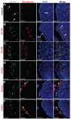
Fluorescent microscope images showing the FAE of PPs that had been stained with lectin UEA I (a marker for alpha-L-fucose residue on M cells) after 4 hours of oral administration of different sizes of fluorescent thiol-organosilica particles. (A) Control, M cells were scattered in the FAE, and alpha-L-fucose residue was expressed on the surface and upper cytoplasm of M cells. PP after administration of (B) Rh100 particles, (C) Rh180 particles, (D) Rh365 particles, (E) Rh745 particles, and (F) Rh925 particles. The expression of alpha-L-fucose residue on the surface and cytoplasm of M cells increased more in animals that had been administered Rh745 or Rh925 than in animals that had been administered the smaller particles Rh100, Rh180, or Rh365 or the control. Note: Scale bar = 50 μm. Abbreviations: FAE, follicle-associated epithelium; PPs, Peyer’s patches; Rh, rhodamine B; SED, subepithelial dome; UEA I, Ulex europaeus agglutinin I.
Figure 3
Figure 3
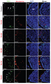
Fluorescent microscope images showing the SED of PPs stained with CD11b (Mac 1α chain) antibody (CD11b) (a marker for macrophages) after 4 hours of oral administration of different sizes of fluorescent thiol-organosilica particles. (A) Control, macrophages scattered in the SEDs of PPs. PPs after administration of (B) Rh100 particles, (C) Rh180 particles, and (D) Rh365 particles; there was co-localization of particles and Cd11b+ macrophages, and the number of CD11b+ macrophages increased compared with the control. PPs after administration of (E) Rh745 particles and (F) Rh925 particles; the number of CD11b+ macrophages were similar to that of the control. CD11b+ macrophages aggregated under the FAE of PPs after administration of all particle sizes. Note: Scale bar = 50 μm. Abbreviations: FAE, follicle associated epithelium; PPs, Peyer’s patches; Rh, rhodamine B; SED, subepithelial dome.
Figure 4
Figure 4
Fluorescent microscope images showing the SED of PPs stained with 33D1 antibody (a marker for DCs) after 4 hours of oral administration of differently sized fluorescent thiol-organosilica particles. (A) Control, 33D1+ DCs scattered in the SED of the PPs. PPs after administration of (B) Rh100 particles, (C) Rh180 particles, and (D) Rh365 particles; there was no co-localization of particles and 33D1+ DCs. The number of 33D1+ DCs was similar to the control. PPs after administration of (E) Rh745 particles and (F) Rh925 particles; the number of 33D1+ DCs increased when compared with the control. 33D1+ DCs aggregated under the FAE of the PPs after administration of all particle sizes. Note: Scale bar = 50 μm. Abbreviations: DCs, dendritic cells; FAE, follicle associated epithelium; PPs, Peyer’s patches; Rh, rhodamine B, SED, subepithelial dome.
Figure 5
Figure 5
Fluorescent microscope images showing PPs at high magnifications after 4 hours of oral administration of Rh100 fluorescently labeled thiol-organosilica particles. (A) CD11b+ macrophages containing particles scattered in the SED of the PPs and aggregated under the FAE of the PPs; large numbers of particles co-localized with CD11b+ macrophages. (B) 33D1+ DCs scattered in the SED of the PPs; there was little co-localization of the particles with 33D1+ DCs. Note: Scale bar = 25 μm. Abbreviations: DCs, dendritic cells; FAE, follicle associated epithelium; PPs, Peyer’s patches; Rh, rhodamine B; SED, subepithelial dome.
Figure 6
Figure 6
Fluorescent microscope images showing the SED of PPs stained with IgA antibody after 4 hours of oral administration of different sizes of fluorescently labeled thiolorganosilica particles. (A) Control, IgA+ cells scattered in the SED of the PPs. PPs after administration of (B) Rh100 particles, (C) Rh180 particles, and (D) Rh365 particles; there was no co-localization of the particles and IgA+ cells. The number of IgA+ cells in animals treated with the smaller particles (Rh100, Rh180, and Rh365) increased compared with the control. PPs after administration of (E) Rh745 particles and (F) Rh925 particles; the number of IgA+ cells were similar to that of the control, but IgA expression on the surface of M cells increased when compared with the smaller-sized particles or the control (white quadrates and enlarged parts of merged (E) and (F)). Notes: Scale bar = (AF), 50 μm; enlarged sections of merged (E) and (F), 10 μm. Abbreviations: DCs, dendritic cells; FAE, follicle associated epithelium; IgA, immunoglobulin A; PPs, Peyer’s patches; Rh, rhodamine B; SED, subepithelial dome.
Figure 7
Figure 7
Fluorescent microscope images showing intestinal lumen (Lu) stained with IgA antibody after 4 hours of oral administration of fluorescently labeled Rh100 or Rh925 thiol-organosilica particles. (A) Control, IgA+ cells (bold arrows) in the villi (V) lamina propria; and (B) Rh100 particles inside the intestinal lumen; no IgA expression inside the lumen was observed, similar to the control. (C) The Rh925 particles inside the intestinal lumen; IgA proteins that were secreted into the lumen and the leukocyte (arrow heads) number increased when compared with that of Rh100-administered animals. Note: Scale bar = 50 μm. Abbreviations: IgA, immunoglobulin A; Lu, lumen; Rh, rhodamine B; V, villi.
Figure 8
Figure 8
Quantitative analysis of the nanoimmunoresponse of immune cells from PPs 4 hours after oral administration of different-sized, fluorescently labeled, thiolorganosilica particles. Immune cells of PPs were counted in 0.154 ± 0.004 mm2 sections of the SEDs of PPs. The uptake of smaller particles (Rh100) increased the number of CD11b+ macrophages and IgA+ cells two- and fourfold, respectively, when compared with larger particles (Rh925) or five- and sevenfold, respectively, when compared with the control. In contrast, mechanical adhesion of larger particles increased the number of 33D1+ DCs twofold when compared with smaller particles or the control. Note: Bars represented means ± standard deviations (n = 20). Abbreviations: DCs, dendritic cells; IgA, immunoglobulin A; PPs, Peyer’s patches; Rh, rhodamine B; SED, subepithelial dome.
Figure 9
Figure 9
Western blotting analysis of IgA induction after 4 hours of oral administration of Rh100 or Rh925 particles. Western blotting analysis of IgA induction in (A) the bile (5 μg/each lane), (B) the intestinal secretions (10 μg/each lane), and (C) in PP homogenates (15 μg/each lane). Smaller thiol-organosilica particles (Rh100) induced IgA in both PP homogenates and bile secretions when compared with larger particles (Rh925); however, the opposite was true for intestinal secretions. Abbreviations: IgA, immunoglobulin A; PPs, Peyer’s patches; Rh, rhodamine B.
Figure 10
Figure 10
Densitometric analysis of IgA induction in the bile, intestinal secretions and PP homogenates after 4 hours of oral administration of Rh100 or Rh925 particles. Note: Bars represent means ± standard deviations (n = 4). Abbreviations: IgA, immunoglobulin A; PPs, Peyer’s patches; Rh, rhodamine B.

Similar articles

Cited by

References

    1. Salman HH, Irache JM, Gamazo C. Immunoadjuvant capacity of flagellin and mannosamine-coated poly(anhydride) nanoparticles in oral vaccination. Vaccine. 2009;27(35):4784–4790. - PubMed
    1. Salman HH, Gamazo C, Agüeros M, Irache JM. Bioadhesive capacity and immunoadjuvant properties of thiamine-coated nanoparticles. Vaccine. 2007;25(48):8123–8132. - PubMed
    1. Chabot S, Wagner JS, Farrant S, Neutra MR. TLRs regulate the gatekeeping functions of the intestinal follicle-associated epithelium. J Immun. 2006;176(7):4275–4283. - PubMed
    1. Shreedhar VK, Kelsall BL, Neutra MR. Cholera toxin induces migration of dendritic cells from the subepithelial dome region to T- and B-Cell areas of Peyer’s patches. Infec Immun. 2003;71(1):504–509. - PMC - PubMed
    1. Semete B, Booysen LI, Kalombo L, et al. In vivo uptake and acute immune response to orally administered chitosan and PEG coated PLGA nanoparticles. Toxicol Appl Pharmacol. 2010;249(2):158–165. - PubMed

Publication types

MeSH terms