. 2012 May;2(2):87-109.
doi: 10.1159/000335675. Epub 2012 Feb 7.

Mitochondria and Oxidative Stress in the Cardiorenal Metabolic Syndrome

Affiliations

Mitochondria and Oxidative Stress in the Cardiorenal Metabolic Syndrome

Annayya R Aroor et al. Cardiorenal Med. 2012 May.

Abstract

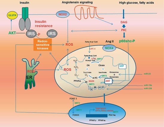
Mitochondria play a fundamental role in the maintenance of normal structure, function, and survival of tissues. There is considerable evidence for mitochondrial dysfunction in association with metabolic diseases including insulin resistance, obesity, diabetes, and the cardiorenal metabolic syndrome. The phenomenon of reactive oxygen species (ROS)-induced ROS release through interactions between cytosolic and mitochondrial oxidative stress contributes to a vicious cycle of enhanced oxidative stress and mitochondrial dysfunction. Activation of the cytosolic and mitochondrial NADPH oxidase system, impairment of the mitochondrial electron transport, activation of p66shc pathway-targeting mitochondria, endoplasmic reticular stress, and activation of the mammalian target of the rapamycin-S6 kinase pathway underlie dysregulation of mitochondrial dynamics and promote mitochondrial oxidative stress. These processes are further modulated by acetyltransferases including sirtuin 1 and sirtuin 3, the former regulating nuclear acetylation and the latter regulating mitochondrial acetylation. The regulation of mitochondrial functions by microRNAs forms an additional layer of molecular control of mitochondrial oxidative stress. Alcohol further exacerbates mitochondrial oxidative stress induced by overnutrition and promotes the development of metabolic diseases.


Figures

Fig. 1
Fig. 1
Sources and consequences of mitochondrial oxidative stress and its relationship to insulin resistance in the CRS. ROS-induced ROS release results from several pathways, including hyperglycemia and lipid-induced activation of PKC, Ang II-mediated activation of cytosolic (NOX2) and mitochondrial (NOX) NADPH oxidase, activation of p66shc, ER stress, and dysregulation of nuclear and mitochondrial transcriptional response. The activation of redox-sensitive kinases induces insulin resistance through increased phosphorylation of serine residues in IRS proteins, which in turn suppresses insulin metabolic signaling.
Fig. 2
Fig. 2
Immunostaining of mitochondrial complex IV-1 in ZO and ZL rats. Representative left ventricular sections immunostained for mitochondrial complex IV-1 of treated and untreated ZL and ZO rats. The increased level of complex IV-1 immunostaining in the ZO-C myocardium indicates an increased mitochondrial number compared with all other groups. The bar graph shows the quantification of converted signal intensities of complex IV-1 protein. ZL-C = Zucker lean-control; ZL-N = Zucker lean-nebivolol; ZO-C = Zucker obese-control; ZO-N = Zucker obese-nebivolol. * p < 0.05 vs. ZL-C; † p < 0.05 vs. ZO-C. Figure used with permission from Zhou et al. [172].
Fig. 3
Fig. 3
miRNA profile in the heart of ZL and ZO rats. The miRNA was isolated with an mirVana miRNA Isolation Kit (Ambion Inc.) from fresh-frozen tissues (n = 3 for each group), and was labeled with a FlashTag™ Biotin HSR RNA Labeling Kit. An Affymetrix miRNA GeneChip was used for this study (46,228 probes comprising 7,815 probe sets, including controls). The probes on this chip are derived from the Sanger miRBase miRNA database v11 (April 15, 2008, http://microrna.sanger.ac.uk) [173]. Data analysis was performed by miRNA QC tool and Significance Analysis of Microarrays (SAM) software [174]. LV = Left ventricle. * p < 0.05 for ZL versus ZO.

References

    1. Ren J, Pulakat L, Whaley-Connell A, Sowers JR. Mitochondrial biogenesis in the metabolic syndrome and cardiovascular disease. J Mol Med. 2010;88:993–1001. - PMC - PubMed
    1. Picard M, Taivassalo T, Gouspillou G, Hepple RT. Mitochondria: isolation, structure and function. J Physiol. 2011;589:4413–4421. - PMC - PubMed
    1. Palmer CS, Osellame LD, Stojanovski D, Ryan MT. The regulation of mitochondrial morphology: intricate mechanisms and dynamic machinery. Cell Signal. 2011;23:1534–1545. - PubMed
    1. Rabøl R, Højberg PM, Almdal T, Boushel R, Haugaard SB, Madsbad S, Dela F. Effect of hyperglycemia on mitochondrial respiration in type 2 diabetes. J Clin Endocrinol Metab. 2009;94:1372–1378. - PubMed
    1. Garcia-Roves PM. Mitochondrial pathophysiology and type 2 diabetes mellitus. Arch Physiol Biochem. 2011;117:177–187. - PubMed

LinkOut - more resources