

doi: 10.1155/2012/302858.
Epub 2012 Apr 26.
Early detection of ovarian cancer with conventional and contrast-enhanced transvaginal sonography: recent advances and potential improvements
Affiliations
- PMID: 22619674
- PMCID: PMC3351123
- DOI: 10.1155/2012/302858
Item in Clipboard
Early detection of ovarian cancer with conventional and contrast-enhanced transvaginal sonography: recent advances and potential improvements
J Oncol.
2012.
Abstract
Recently, there have been several major technical advances in the sonographic diagnosis of ovarian cancer in its early stages. These include improved assessment of tumor morphology with transvaginal sonography (TVS), and detection and characterization of tumor neovascularity with transvaginal color Doppler sonography (TV-CDS) and contrast-enhanced transvaginal sonography (CE-TVS). This paper will discuss and illustrate these improvements and describe how they enhance detection of early-stage ovarian cancer. Our initial experience with parametric mapping of CE-TVS will also be mentioned.
Figures

Morphologic signs of malignancy with histopathologic correlation on TVS in various histologic types of stage 1A ovarian cancer.

2D CDS of showing flow within papillary excrescence within a papillary cystadenofibroma.

3D TV-CDS of papillary excrescences within a papillary serous cystadenoma.

3D TV-CDS of papillary cystadenocarcinoma showing multiplanar reconstruction (MPR) images.

CE-TVS of a benign fibroma.

CE-TVS of serous cystadenoma with mural nodules.

CE-TVS of borderline mucinous (intestinal) cystadenocarcinoma.

CE-TVS of stage I papillary serous cystadenocarcinoma.

Box graph of contrast-enhanced parameters. While there is no difference in time of peak (T wash-in), there are significant differences in peak enhancement, wash-out time and vascularity ((b), (c); (d)) from [1].

Sensitivities and specificities of maximum enhancement, wash-in, wash-out and area under curve (AUC). Maximum enhancement, wash-out and AUC had greatest accuracy.

Receiver operator characteristerics for (a) wash-in, (b) maximum enhancement, (c) wash-out, and (d) area under curve.
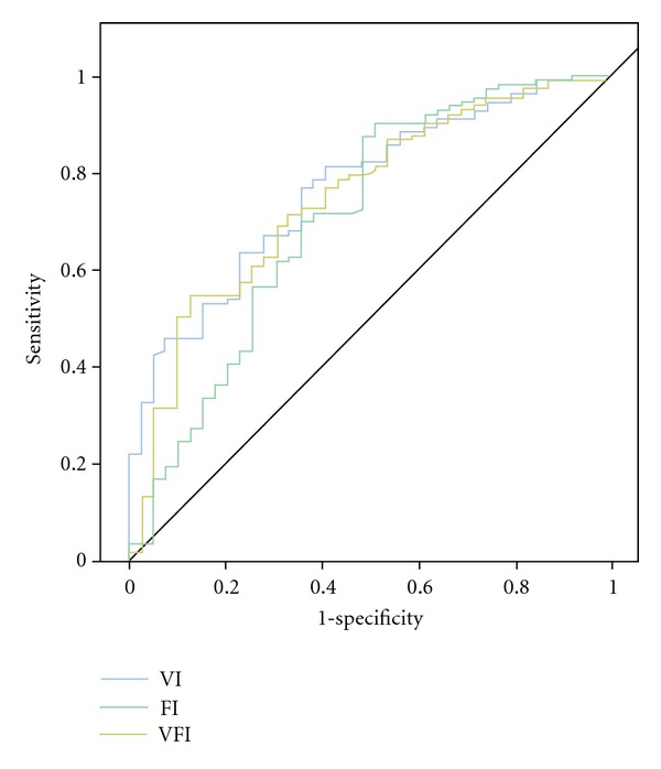
Receiver operator characteristic of various parameters showing cutoff points for vascular index (VI), flow index (FI), and vascular flow index (VFI) (from [2]).

Relative accuracy (sensitivity and specificity of enhancement kinetic parameters) of various techniques using predetermined cutoff points of: 2D VFI (>0.4), 3D VFI (>0.5), CE-TVS (max >17.2 dB), CE-TVS ((1/2)T
wo > 41 sec), CE-TVS (AUC > 787 s−1).
Similar articles
-
Transvaginal color Doppler sonography and conventional sonography in the preoperative assessment of adnexal masses.J Clin Ultrasound. 1997 Jun;25(5):217-25. doi: 10.1002/(sici)1097-0096(199706)25:5<217::aid-jcu1>3.0.co;2-g. J Clin Ultrasound. 1997. PMID: 9314102
-
Advances in sonographic detection of ovarian cancer: depiction of tumor neovascularity with microbubbles.AJR Am J Roentgenol. 2010 Feb;194(2):343-8. doi: 10.2214/AJR.09.3446. AJR Am J Roentgenol. 2010. PMID: 20093594 Review.
-
The detection of stage I ovarian cancer by three-dimensional sonography and power Doppler.Gynecol Oncol. 2003 Aug;90(2):258-64. doi: 10.1016/s0090-8258(03)00205-1. Gynecol Oncol. 2003. PMID: 12893185
-
Ovarian cancer screening.Cancer. 1995 Nov 15;76(10 Suppl):2086-91. doi: 10.1002/1097-0142(19951115)76:10+<2086::aid-cncr2820761330>3.0.co;2-l. Cancer. 1995. PMID: 8635005 Review.
-
The efficacy of transvaginal sonographic screening in asymptomatic women at risk for ovarian cancer.Gynecol Oncol. 2000 Jun;77(3):350-6. doi: 10.1006/gyno.2000.5816. Gynecol Oncol. 2000. PMID: 10831341 Clinical Trial.
Cited by
-
Clinicopathological Characteristics and Prognostic Factors in Ovarian Metastases from Right- and Left-Sided Colorectal Cancer.Curr Oncol. 2021 Aug 3;28(4):2914-2927. doi: 10.3390/curroncol28040255. Curr Oncol. 2021. PMID: 34436021 Free PMC article.
-
Risk Algorithm Using Serial Biomarker Measurements Doubles the Number of Screen-Detected Cancers Compared With a Single-Threshold Rule in the United Kingdom Collaborative Trial of Ovarian Cancer Screening.J Clin Oncol. 2015 Jun 20;33(18):2062-71. doi: 10.1200/JCO.2014.59.4945. Epub 2015 May 11. J Clin Oncol. 2015. PMID: 25964255 Free PMC article.
-
Radiologic-Histopathologic Correlation of Transvaginal US and Risk-reducing Salpingo-oophorectomy for Women at High Risk for Tubo-ovarian Carcinoma.Radiol Imaging Cancer. 2020 Nov 13;2(6):e190086. doi: 10.1148/rycan.2020190086. eCollection 2020 Nov. Radiol Imaging Cancer. 2020. PMID: 33778746 Free PMC article.
-
Urine Biomarkers for the Early Detection of Ovarian Cancer - Are We There Yet?Biomark Cancer. 2019 Feb 26;11:1179299X19830977. doi: 10.1177/1179299X19830977. eCollection 2019. Biomark Cancer. 2019. PMID: 30833816 Free PMC article. Review.
-
Transvaginal ultrasonography in ovarian cancer screening: current perspectives.Int J Womens Health. 2013 Dec 20;6:25-33. doi: 10.2147/IJWH.S38347. Int J Womens Health. 2013. PMID: 24379701 Free PMC article. Review.
References
-
- Hirai M, Hirai Y, Tschida T, et al. Comparison of sonographic findings and histopathologic types between patients with normal and elevated serum cancer antigen 125 levels. Journal of Ultrasound in Medicine. 2011;30(7):943–952. - PubMed
-
- Jinawath N, Shih I. Biology and pathology of ovarian cancer. In: Briston R, Armstrong D, editors. Early Diagnosis and Treatment of Cancer. Ovarian Cancer. Philadelphia, Pa, USA: Saunders; 2011. pp. 17–32.
-
- Bazot M, Daraï E, Nassar-Slaba J, Lafont C, Thomassin-Naggara I. Value of magnetic resonance imaging for the diagnosis of ovarian tumors: a review. Journal of Computer Assisted Tomography. 2008;32(5):712–723. - PubMed
-
- Risum S, Hogdall C, Loft A, et al. The diagnostic value of PET/CT for primary ovarian cancer: a prospective study. Gynecologic Oncology. 2007;105(1):145–149. - PubMed
Grants and funding
LinkOut - more resources
Full Text Sources
Medical
