. 2012 May 23;14(3):R85.
doi: 10.1186/bcr3200.

JMJD6 is a driver of cellular proliferation and motility and a marker of poor prognosis in breast cancer

Affiliations

JMJD6 is a driver of cellular proliferation and motility and a marker of poor prognosis in breast cancer

Yi Fang Lee et al. Breast Cancer Res. .

Erratum in

Abstract

Introduction: We developed an analytic strategy that correlates gene expression and clinical outcomes as a means to identify novel candidate oncogenes operative in breast cancer. This analysis, followed by functional characterization, resulted in the identification of Jumonji Domain Containing 6 (JMJD6) protein as a novel driver of oncogenic properties in breast cancer.

Methods: Through microarray informatics, Cox proportional hazards regression was used to analyze the correlation between gene expression and distant metastasis-free survival (DMFS) of patients in 14 independent breast cancer cohorts. JMJD6 emerged as a top candidate gene robustly associated with poor patient survival. Immunohistochemistry, siRNA-mediated silencing, and forced overexpression of JMJD6 in cell-based assays elucidated molecular mechanisms of JMJD6 action in breast cancer progression and shed light on the clinical breast cancer subtypes relevant to JMJD6 action.

Results: JMJD6 was expressed at highest levels in tumors associated with worse outcomes, including ER- and basal-like, Claudin-low, Her2-enriched, and ER+ Luminal B tumors. High nuclear JMJD6 protein was associated with ER negativity, advanced grade, and poor differentiation in tissue microarrays. Separation of ER+/LN- patients that received endocrine monotherapy indicated that JMJD6 is predictive of poor outcome in treatment-specific subgroups. In breast cancer cell lines, loss of JMJD6 consistently resulted in suppressed proliferation but not apoptosis, whereas forced stable overexpression increased growth. In addition, knockdown of JMJD6 in invasive cell lines, such as MDA-MB231, decreased motility and invasion, whereas overexpression in MCF-7 cells slightly promoted motility but did not confer invasive growth. Microarray analysis showed that the most significant transcriptional changes occurred in cell-proliferation genes and genes of the TGF-β tumor-suppressor pathway. High proliferation was characterized by constitutively high cyclin E protein levels. The inverse relation of JMJD6 expression with TGF-β2 could be extrapolated to the breast cancer cohorts, suggesting that JMJD6 may affect similar pathways in primary breast cancer.

Conclusions: JMJD6 is a novel biomarker of tumor aggressiveness with functional implications in breast cancer growth and migration.


Figures

Figure 1
Figure 1
High JMJD6 mRNA expression is associated with poor survival outcome. Cox regression analysis of JMJD6 expression and distant metastasis-free survival of patients was initially performed in 14 microarray expression datasets independently and then subsequently on a "Super Cohort" (SC) comprising 15 Affymetrix array-based cohorts, including a subset of the 14 cohorts used initially. Hazard ratios and their 95% confidence intervals are shown in the forest plot. Datasets are listed on the Y-axis; all platforms are from Affymetrix array series, with the exception of NKI from Agilent. The 95% confidence intervals are indicated by the error bars, and the cohort size is shown in parentheses. *P ≤ 0.05; **P ≤ 0.01; ***P ≤ 0.005.
Figure 2
Figure 2
JMJD6 expression and prognosis in intrinsic subtypes of breast cancer. Box plots of JMJD6 expression demonstrate (A) higher JMJD6 probeset intensity (log2) in ER- tumors compared with ER+ tumors, with P < 0.001 by Mann-Whitney Rank Sum test; and (B) highest JMJD6 probeset intensity (log2) in claudin-low and basal subtypes, followed by HER2-enriched and LumB subtypes, and lowest in LumA subtypes. The Dunn method was used to perform pairwise multiple comparison among the subtypes. *P < 0.05 between a pair of subtypes. Dots of boxplots (A, B) represent outliers in the 90th and 10th percentiles. Kaplan-Meier survival curves based on below-median (low) and above-median (high) JMJD6 expression are shown for (C) ER+ patients, (D) patients of LumA subtype, (E) patients of LumB subtype, and (F) ER+, tamoxifen-treated patients. The numbers in the parentheses equal patient numbers in each group, and the log rank P value is indicated at the bottom left of each figure.
Figure 3
Figure 3
Nuclear JMJD6 protein expression is associated with high-grade breast tumors. Representative breast cancer cores from tissue microarray that were stained with JMJD6 antibody are shown. Low-grade (left panel), intermediate-grade (middle panel), and high-grade breast tumors (right panel) are shown (magnification, ×60). The table in the figure shows univariate analysis of JMJD6 and clinical parameters.
Figure 4
Figure 4
Expression of JMJD6 enhances proliferation. (A) WST-1 assays using MCF-7-J1-OE clones (J1-C2, J1-C3, and J1-C7) showed increased proliferation over vector control cells (Vec). SiRNA-mediated knockdown of JMJD6 and resultant decrease in proliferation is shown in (B) MCF-7 and MDA-MB231. Four individual siRNAs (A, B, C, and D) were used: Sc, scrambled siRNA; and NT, non-transfected cells served as control. Inset in each panel shows protein levels of JMJD6 after siRNA transfection. β-actin immunoblots show equal protein loading in all lanes.
Figure 5
Figure 5
JMJD6 promotes motility in breast cancer cell lines. (A) Scatter phenotype was scored based on three levels of scattering (compact, loose, scatter) (upper panel). Bar graphs represent quantification of scattering of colonies by using the Student t test (lower panel). MCF-7 J1 OE clones had a higher percentage of scattered or loosely scattered clones than did the control Vec cells. (B) Wound-healing assay showed that MCF-7 J1 clones displayed higher motility in comparison to Vec (upper panel); quantification of wound closure as a ratio to the Vec is shown in the lower panel. *P ≤ 0.05 by Student t test in comparison to Vec control. (C) Boyden-chamber assay results of MDA-MB231 cells with JMJD6 siRNA-mediated knockdown were normalized to fold change in proliferation at Day 2 of WST-1 measurement. Two of three siRNA knockdowns of JMJD6 showed significantly decreased motility, and all siRNA knockdowns of JMJD6 showed decreased invasiveness. *P ≤ 0.05; **P ≤ 0.001 by Student's t- test when compared with Sc siRNA.
Figure 6
Figure 6
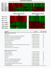
IPA analysis of JMJD6-regulated genes showed enrichment in cell-cycle function. (A) The heatmap (top panel) shows that hierarchic clustering of the genes changed after siRNA knockdown and overexpression of JMJD6. The sample labels are on the top of the heat map. Two clusters representing JMJD6-induced and JMJD6-repressed genes were selected (middle heat-map panel) and subjected to IPA analysis. (B) Table shows a list of the top 10 functions that are enriched in cells with altered levels of JMJD6.
Figure 7
Figure 7
JMJD6 represses TGF-β2 expression. RT-qPCR assays of TGF-βs after JMJD6 siRNA-mediated knockdown are shown in (A) MCF-7 and (B) MDA-MB231. Consistently, TGF-β2 and TGF-β1 mRNA levels were upregulated on JMJD6 knockdown. (C) Immunoblots showed that the amount of secreted TGF-β2 protein in conditioned media was higher after JMJD6 siRNA-mediated knockdown in MCF-7 and MDA-MB231. (D) RT-qPCR assay showed that MCF7 J1 OE clones had a lower level of TGF-β1 and TGF-β2 transcripts than did the Vec cells. (E) Western blot showed a dramatic decrease in secreted TGF-β2 protein, in the MCF-7 J1 clones, as compared with the Vec cells. NS, a Ponceau-stained blot of the conditioned media to demonstrate protein loading. Student's t- test was performed by using scrambled siRNA versus JMJD6 siRNA and Vec cells versus MCF-7J1 clones. *P ≤ 0.05; **P ≤ 0.005.
Figure 8
Figure 8
JMJD6 expression affects SMAD phosphorylation. Immunoblots show that the levels of total SMAD2/3, phosphorylated SMAD2, and phosphorylated SMAD3 are decreased in MCF-7 J1-OE clones and increased in JMJD6 siRNA-mediated knockdown in MCF-7 and MDA-MB231. The numbers next to the SMAD2P/3P denote the expected sites of phosphorylation detected by the antibodies. NS, nonspecific bands from the blots, which indicate equal loading of the protein lysates.
Figure 9
Figure 9
TGF-β2 inhibits proliferation in MCF-7. (A) Immunoblots showed that treatment of MCF-7 by recombinant human (rh) TGF-β2 (5 ng/ml) resulted in enhanced Smad2 phosphorylation, which could be effectively nullified by SB431542. (B) Treatment of JMJD6 siRNA-transfected MCF-7 cells with SB431542 (10 μM) resulted in rescue of the proliferation defect. Ratio of OD at Day 3 to Day 1 in JMJD6 knockdown cells was normalized to the scrambled siRNA control. *P ≤ 0.05; **P ≤ 0.005 between DMSO and SB431542 treatments. (C) Treatment of MCF-7 J1-OE and Vec cells with rhTGF-β2 resulted in decreased proliferation, as measured by WST-1 assay. **P ≤ 0.001 between BSA and rhTGF-β2 treatments.
Figure 10
Figure 10
Decrease of cyclin E2 protein level in JMJD6 knockdown. Western blot detection of G1 cell-cycle cyclins is shown. A decrease in cyclin E2 (CCNE2) level with decrease in JMJD6 levels was evident in MCF-7 and MDA-MB231. Cyclin E1 (CCNE1) and cyclin D1 (CCND1) displayed an inconsistent change on JMJD6 siRNA transfection in both MCF-7 and MDA-MB231 cells.
Figure 11
Figure 11
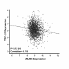
Clinical association of JMJD6 with TGF-β2. Pearson correlation of clinical expression of JMJD6 with TGF-β2 suggests a negative correlation with TGF-β2 (P = 9.13 × 10⁻⁸). Scatterplot shows the normalized JMJD6 expression of each patient sample with the corresponding normalized TGF-β2 expression in the clinical dataset of 2,034 breast cancer patients.

References

    1. Soon WWML, Black MA, Dalmasso C, Chan XB, Pang B, Ong CW, Salto-Tellez M, Desai K, Liu ET. Combined genomic and phenotype screening reveals secretory factor SPINK1 as an invasion and survival factor associated with patient prognosis in breast cancer. EMBO Mol Med. 2011;3:451–464. doi: 10.1002/emmm.201100150. - DOI - PMC - PubMed
    1. Fadok VA, Bratton DL, Rose DM, Pearson A, Ezekewitz RAB, Henson PM. A receptor for phosphatidylserine-specific clearance of apoptotic cells. Nature. 2000;405:85–90. doi: 10.1038/35011084. - DOI - PubMed
    1. Bose J, Gruber A, Helming L, Schiebe S, Wegener I, Hafner M, Beales M, Kontgen F, Lengeling A. The phosphatidylserine receptor has essential functions during embryogenesis but not in apoptotic cell removal. J Biol. 2004;3:15. doi: 10.1186/jbiol10. - DOI - PMC - PubMed
    1. Cui P, Qin B, Liu N, Pan G, Pei D. Nuclear localization of the phosphatidylserine receptor protein via multiple nuclear localization signals. Exp Cell Res. 2004;293:154–163. doi: 10.1016/j.yexcr.2003.09.023. - DOI - PubMed
    1. Chang B, Chen Y, Zhao Y, Bruick RK. JMJD6 Is a histone arginine demethylase. Science. 2007;318:444–447. doi: 10.1126/science.1145801. - DOI - PubMed

Publication types

MeSH terms