. 2012;28(4):320-36.
doi: 10.3109/02656736.2012.680173.

Mild hyperthermia with magnetic resonance-guided high-intensity focused ultrasound for applications in drug delivery

Affiliations

Mild hyperthermia with magnetic resonance-guided high-intensity focused ultrasound for applications in drug delivery

Ari Partanen et al. Int J Hyperthermia. 2012.

Erratum in

  • Int J Hyperthermia. 2012;28(5):473

Abstract

Purpose: Mild hyperthermia (40-45 °C) is a proven adjuvant for radiotherapy and chemotherapy. Magnetic resonance guided high intensity focused ultrasound (MR-HIFU) can non-invasively heat solid tumours under image guidance. Low temperature-sensitive liposomes (LTSLs) release their drug cargo in response to heat (>40 °C) and may improve drug delivery to solid tumours when combined with mild hyperthermia. The objective of this study was to develop and implement a clinically relevant MR-HIFU mild hyperthermia heating algorithm for combination with LTSLs.

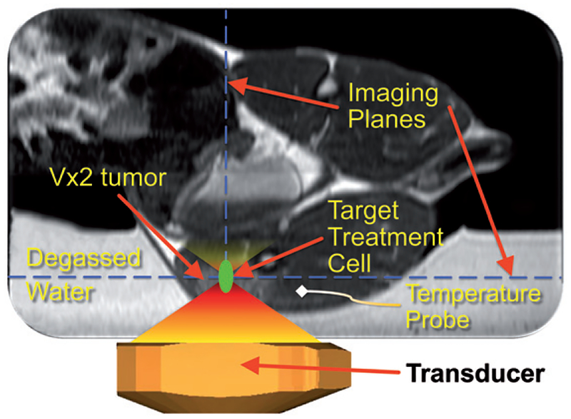
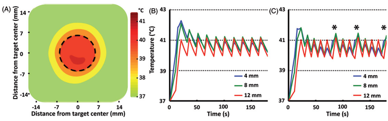
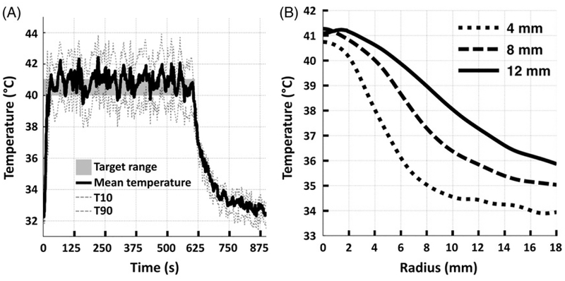
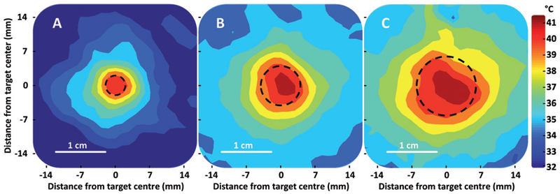
Materials and methods: Sonications were performed with a clinical MR-HIFU platform in a phantom and rabbits bearing VX2 tumours (target = 4-16 mm). A binary control algorithm was used for real-time mild hyperthermia feedback control (target = 40-41 °C). Drug delivery with LTSLs was measured with HPLC. Data were compared to simulation results and analysed for spatial targeting accuracy (offset), temperature accuracy (mean), homogeneity of heating (standard deviation (SD), T10 and T90), and thermal dose (CEM43).

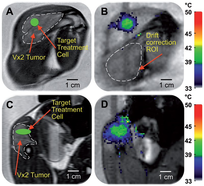
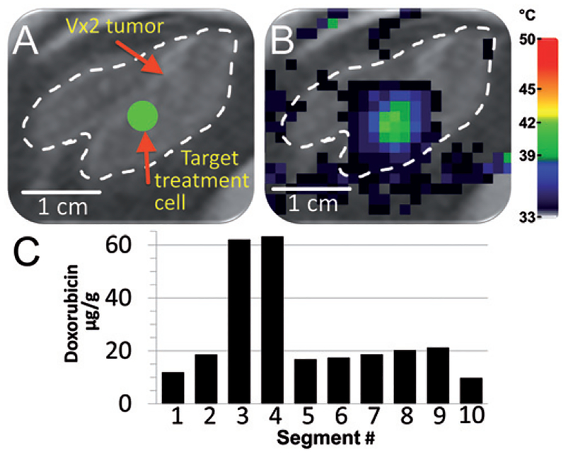
Results: Sonications in a phantom resulted in better temperature control than in vivo. Sonications in VX2 tumours resulted in mean temperatures between 40.4 °C and 41.3 °C with a SD of 1.0-1.5 °C (T10 = 41.7-43.7 °C, T90 = 39.0-39.6 °C), in agreement with simulations. 3D spatial offset was 0.1-3.2 mm in vitro and 0.6-4.8 mm in vivo. Combination of MR-HIFU hyperthermia and LTSLs demonstrated heterogeneous delivery to a partially heated VX2 tumour, as expected.

Conclusions: An MR-HIFU mild hyperthermia heating algorithm was developed, resulting in accurate and homogeneous heating within the targeted region in vitro and in vivo, which is suitable for applications in drug delivery.


Figures

Figure 1.
Figure 1.
Schematic of the experimental MR-HIFU hyperthermia set-up, modified from Ranjan et al. [66]. The sagittal imaging plane is shown, with the rabbit in right lateral decubitus position on top of the HIFU platform and the tumour-bearing right hind limb submerged in degassed water. Baseline reference temperature was obtained using a fibre-optic temperature probe inserted in the thigh muscle near the tumour. The imaging slice positions for the thermometry sequence are outlined with a blue dashed line, and the target region within the tumour is shown as a green circle. Depiction of transducer and HIFU beam propagation are meant to be illustrative.
Figure 2.
Figure 2.
Planning and temperature mapping for mild hyperthermia: (A) VX2 tumour (hyper-intense) was clearly identified (white dashed line) on the proton density-weighted planning images and a target region within the tumour was chosen (green circle). (B) Temperature maps (colour scale) overlaid on planning images (greyscale) during a mild hyperthermia treatment with an 8 mm treatment cell, showing typical temperature distribution after 5 min of heating. Temperature monitoring and control was achieved in the selected target region with an FFE-EPI imaging sequence, utilising the PRFS method for temperature mapping, and by using the mild hyperthermia feedback control algorithm. The ROI used for magnetic drift correction is outlined with a white dashed line. C and D are the sagittal image planes corresponding to A and B, respectively.
Figure 3.
Figure 3.
Mild hyperthermia feedback schematic. (A) During heat-up, sonication cycles through all heat-up trajectories. Once the criteria for every heat-up trajectory are met, heating is paused. When temperature in one of the monitored subtrajectories drops below the lower limit, sonication resumes on that subtrajectory until the upper limit is reached. The cycle of ‘wait’ and ‘maintain’ subtrajectories is repeated until the end of treatment. (B) The schematic demonstrates the flexibility of the binary feedback control algorithm. The algorithm sequentially heats from the innermost to the outermost subtrajectory during heat-up. After the outermost subtrajectory has been heated sufficiently, the algorithm pauses sonication in a ‘wait subtrajectory’. When temperature in one of the subtrajectories decreases below a predefined range, the algorithm is able to heat that subtrajectory specifically.
Figure 4.
Figure 4.
(A) The mean temperature within an 8mm treatment cell over a 10 min sonication + additional 5 min monitoring in vivo. Uncorrected (grey line) and corrected (black line) temperatures clearly demonstrate the effect of B0 magnetic field drift. (B) The total baseline temperature drift over 15 min from the same sonication as in A resulted in a change of 3°C.
Figure 5.
Figure 5.
(A) Time-averaged spatial temperature distribution for a 12mm treatment cell in silico (coronal plane) for 10 min mild hyperthermia with normal perfusion (1 mL/mL/min). Treatment cell is outlined in black dashed line.(B, C) Simulated mean temperature along 4, 8 and 12 mm subtrajectories at two different perfusion levels (B, normal perfusion (1 mL/mL/min) and C, high perfusion (2 mL/mL/min). Only at high perfusion level was it necessary to heat subtrajectories other than 12mm after initial heat-up – notice the heating of the 4mm subtrajectory at 80, 120 and 170 s (marked with asterisks in C).
Figure 6.
Figure 6.
(A) Representative examples of mean (solid), T10, and T90 (dashed) temperatures within an 8mm treatment cell over a 10 min sonication in vivo. Target temperature range is indicated as a grey box. (B) Representative examples of time-averaged mean temperature radial line profiles centred on the treatment cell for 4 mm, 8mm, and 12 mm treatment cells.
Figure 7.
Figure 7.
Representative examples of time-averaged spatial temperature distributions for 4 mm, 8 mm, and 12 mm treatment cells in vivo (coronal plane). The treatment cell is outlined in black dashed line.
Figure 8.
Figure 8.
Demonstration of heterogeneous drug delivery. (A) VX2 tumour was clearly identified (white dashed line) on the proton density-weighted planning images and a target region within the tumour was chosen (green circle). (B) Temperature maps (colour scale) overlaid on planning images (greyscale) during a mild hyperthermia treatment with a 4mm treatment cell, showing typical temperature distribution after 1 min of heating. (C) Doxorubicin concentration in tumour segments was determined by HPLC. Note the higher drug concentration in segments 3 and 4.

References

    1. Viglianti BL, Stauffer P, Repasky E, Jones E, Vujaskovic Z, Dewhirst MW. Hyperthermia In: Hong W, Bast R Jr, Hait W, Kufe DW, Holland JF, Pollock RE, et al. , editors. Holland Frei Cancer Medicine. Shelton, CT: Peoples Medical Publishing House-USA; 2010. pp 528–540.
    1. Issels RD, Lindner LH, Verweij J, Wust P, Reichardt P, Schem BC, et al. Neo-adjuvant chemotherapy alone or with regional hyperthermia for localised high-risk soft-tissue sarcoma: A randomised phase 3 multicentre study. Lancet Oncol 2010;11:561–570. - PMC - PubMed
    1. Wood BJ, Ramkaransingh JR, Fojo T, Walther MM, Libutti SK. Percutaneous tumor ablation with radiofrequency. Cancer 2002;94:443–451. - PMC - PubMed
    1. Meshorer A, Prionas SD, Fajardo LF, Meyer JL, Hahn GM, Martinez AA. The effects of hyperthermia on normal mesenchymal tissues. Application of a histologic grading system. Arch Pathol Lab Med 1983;107:328–334. - PubMed
    1. Köhler MO, Mougenot C, Quesson B, Enholm J, Le Bail B, Laurent C, et al. Volumetric HIFU ablation under 3D guidance of rapid MRI thermometry. Med Phys 2009;36:3521–3535. - PubMed

Publication types

MeSH terms