. 2012 Jun;122(6):2006-17.
doi: 10.1172/JCI57513. Epub 2012 May 24.

Blood flow reprograms lymphatic vessels to blood vessels

Affiliations

Blood flow reprograms lymphatic vessels to blood vessels

Chiu-Yu Chen et al. J Clin Invest. 2012 Jun.

Erratum in

  • J Clin Invest. 2012 Jul 2;122(7):2702. Vincente, Andres [corrected to Vicente, Andres]

Abstract

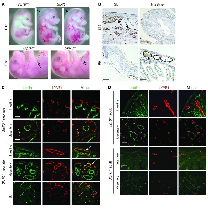
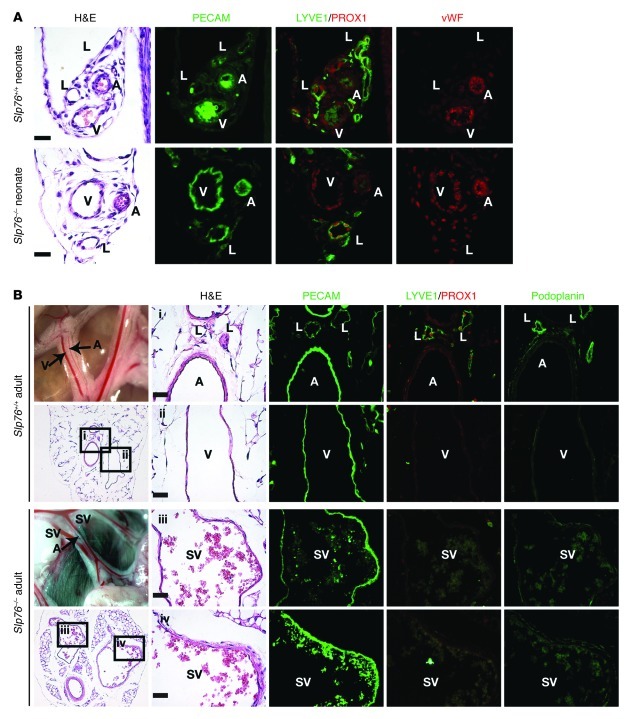
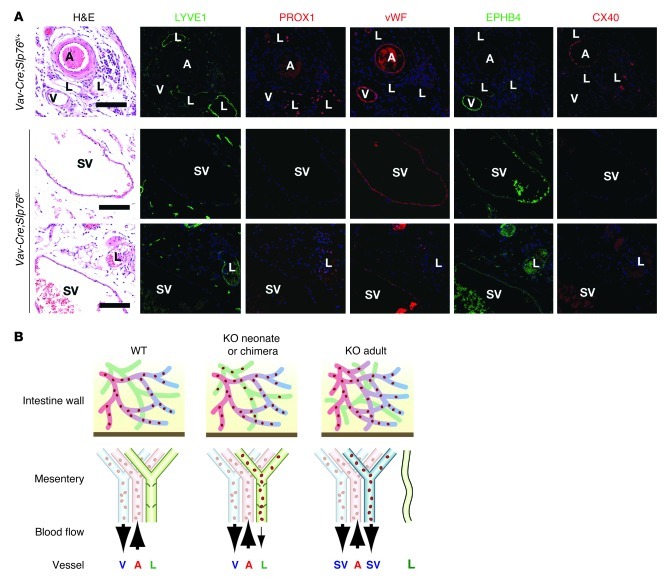
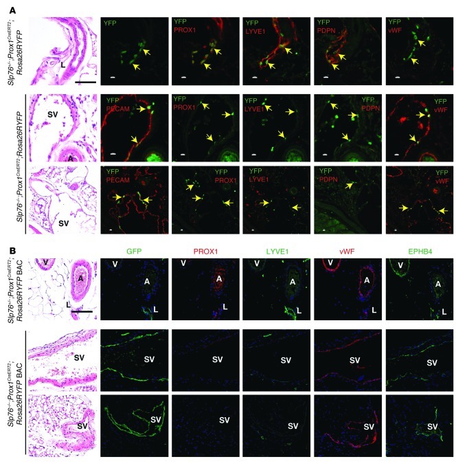
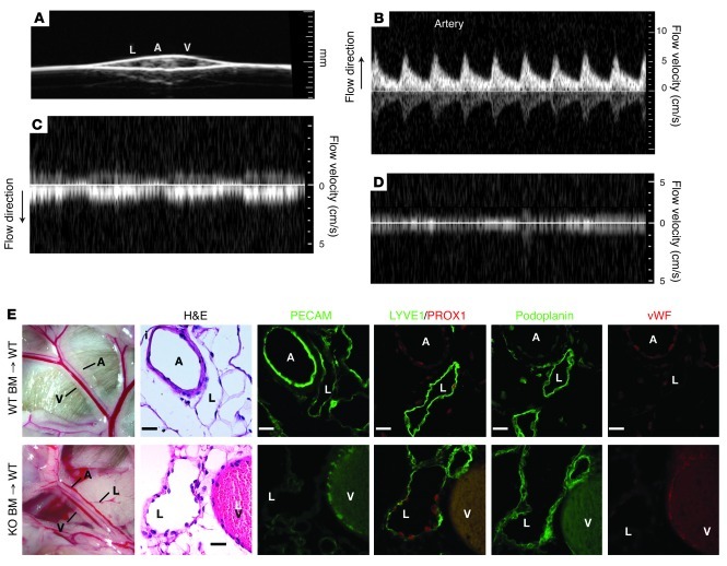
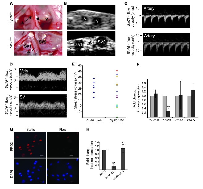
Human vascular malformations cause disease as a result of changes in blood flow and vascular hemodynamic forces. Although the genetic mutations that underlie the formation of many human vascular malformations are known, the extent to which abnormal blood flow can subsequently influence the vascular genetic program and natural history is not. Loss of the SH2 domain-containing leukocyte protein of 76 kDa (SLP76) resulted in a vascular malformation that directed blood flow through mesenteric lymphatic vessels after birth in mice. Mesenteric vessels in the position of the congenital lymphatic in mature Slp76-null mice lacked lymphatic identity and expressed a marker of blood vessel identity. Genetic lineage tracing demonstrated that this change in vessel identity was the result of lymphatic endothelial cell reprogramming rather than replacement by blood endothelial cells. Exposure of lymphatic vessels to blood in the absence of significant flow did not alter vessel identity in vivo, but lymphatic endothelial cells exposed to similar levels of shear stress ex vivo rapidly lost expression of PROX1, a lymphatic fate-specifying transcription factor. These findings reveal that blood flow can convert lymphatic vessels to blood vessels, demonstrating that hemodynamic forces may reprogram endothelial and vessel identity in cardiovascular diseases associated with abnormal flow.


Figures

Figure 1
Figure 1. Late separation of the blood and lymphatic circulations in SLP76-deficient mice.
(A) Cutaneous edema resolves in Slp76–/– embryos between E15 and E18. Arrows indicate sites of raised skin indicative of edema at E15 (top) and the presence of normal skin folds indicating a lack of edema at E18 (bottom). (B) Blood-filled lymphatics resolve in the skin of Slp76–/– embryos at the same time that they appear in the intestine. LYVE1 staining (brown) identifies lymphatic vessels in the skin and intestine of E15 and neonatal Slp76–/– animals. Arrows indicate blood-filled lymphatics in E15 skin and in neonatal intestine. (C) Functional identification of blood-filled lymphatics in neonatal Slp76–/– animals using intravenous injection of biotinylated lectin. Blood-perfused vessels are identified by FITC-streptavidin binding to biotin-lectin after intravenous injection (green), and lymphatic vessels are visualized by LYVE1 immunostaining (red). Co-stained vessels are present in the intestine and mesentery but not skin of Slp76–/– neonates (arrows). (D) Late resolution of mesenteric and intestinal blood-lymphatic mixing in Slp76–/– mice demonstrated using staining for injected biotin-lectin and LYVE1. Scale bars: 50 μm.
Figure 2
Figure 2. Mesenteric SVs in adult Slp76–/– mice lose lymphatic identity.
(A) Neonatal mesenteric lymphatics in Slp76–/– mice express the lymphatic endothelial molecular markers LYVE1 and PROX1, but not the blood endothelial marker vWF. Shown is antibody staining of serial sections. (B) The mesenteric SVs in adult Slp76–/– mice express PECAM but not LYVE1, PDPN, or PROX1. A, artery; V, vein; L, lymphatic. Scale bars: 20 μm.
Figure 3
Figure 3. Mesenteric SVs in adult Vav-Cre;Slp76fl/– mice lose lymphatic identity and acquire venous identity.
(A) Mesenteric SVs that form in Vav-Cre;Slp76fl/– mice lose expression of lymphatic molecular markers and acquire expression of the blood vessel marker vWF and venous marker EPHB4 but not the arterial marker CX40. Scale bars: 50 μm. (B) Model of vascular remodeling in SLP76-deficient mice. Shown are the vascular anatomy and flow through the intestinal and mesenteric vessels of neonatal wild-type, neonatal Slp76–/– (KO neonate), and mature Slp76–/– (KO adult) animals. SLP76-deficient radiation chimeras (KO chimera) develop a vascular phenotype like that observed in KO neonates. In the wild-type animal, afferent mesenteric blood flow is carried by the mesenteric artery (A, red), while efferent blood and lymph are carried by the mesenteric vein (V, blue) and lymphatic (L, green), respectively. In the KO neonate or KO chimera, blood-lymphatic mixing allows blood to enter the mesenteric lymphatics, but flow is minimal, and lymphatic identity is preserved. In the KO adult, mesenteric lymphatics become incorporated into an arterio-venous shunt that directs efferent blood flow through them (right), a process that produces an SV (blue) with blood vessel identity. Histologic studies reveal the presence of small lymphatic vessels that retain lymphatic identity in mature KO animals.
Figure 4
Figure 4. Genetic lineage tracing demonstrates that the blood endothelial cells lining mesenteric SVs in Slp76–/– mice arise from LECs.
(A) Lineage tracing studies performed using a Prox1CreERT2 knock-in line. Antibody staining of mesentery from 12-week-old Slp76+/–;Prox1CreERT2;Rosa26RYFP animals exposed to tamoxifen as neonates reveals YFP only in PROX1+LYVE1+PDPN+vWFLECs (top). Antibody staining of mesentery from 12-week-old Slp76–/–;Prox1CreERT2;Rosa26RYFP animals exposed to tamoxifen as neonates reveals YFP in PROX1LYVE1PDPNvWF+ blood endothelial cells that line large SVs (bottom). Black scale bars: 50 μm; white scale bars: 10 μm. (B) Lineage tracing studies using a Prox1CreERT2 BAC transgenic line. Studies were performed as described in A using a single tamoxifen injection at P14. Note that with this Prox1CreERT2 line, virtually all the endothelial cells of the SVs shown are YFP+ but PROX1LYVE1PDPNvWF+EPHB4+. Scale bar: 50 μm; applies to all panels in B.
Figure 5
Figure 5. Lymphatic endothelial identity is maintained in the presence of blood in Slp76–/– radiation chimeras.
(A) The mesenteric vessels of Slp76–/– radiation chimeras were visualized using 2D ultrasound. (BD) Pulsed-wave Doppler signals indicative of blood flow detected in the mesenteric artery, vein, and lymphatic are shown. Note that arterial flow is directed opposite to that of venous flow and that arterial flow is pulsatile (B, corresponding to heart rate), while venous flow is phasic (C, corresponding to respiration). No significant signal in either direction was obtained from the lymphatic (D). (E) Blood-filled mesenteric lymphatics retain lymphatic identity in Slp76–/– radiation chimeras. Mesenteric vessels in wild-type lethally irradiated mice reconstituted with Slp76+/+ (+/+) or Slp76–/– (–/–) bone marrow are shown 16 weeks after reconstitution (left). Analysis of serial sections reveals that mesenteric lymphatics in Slp76–/– radiation chimeras that are exposed to blood but not flow retain expression of LYVE1, PROX1, and PDPN and do not express vWF (right). Scale bars: 50 μm.
Figure 6
Figure 6. Fluid shear forces equivalent to those in Slp76–/– mesenteric lymphatics drive loss of PROX1 expression in LECs.
(A) Representative mesenteric vessels in 12-week-old Slp76+/+ and Slp76–/– animals that were studied using Doppler ultrasound. The dilated, blood-containing vein and congenital lymphatic are denoted as SV1 and SV2. (B) The vessels shown in A were visualized using 2D ultrasound. (C and D) The pulsed-wave Doppler signals and measured flow velocity of blood in the vessels indicated in A and B are shown. Note the difference in direction of flow between the arteries and veins or efferent SVs. (E) Calculated shear stresses for Slp76+/+ veins and Slp76–/– SVs. Slp76–/– data points in the same color indicate values of paired SVs from the same mesenteric bundle. (F) LECs exposed to flow downregulate PROX1. LECs were subjected to a shear stress of 20 dynes/cm2 for 8 hours, and the expression of the indicated mRNAs measured using qPCR. n = 7. **P < 0.01. (G) Loss of PROX1 protein in LECs exposed to shear. Anti-PROX1 immunostaining in primary LECs is shown compared with DAPI staining of cell nuclei. (H) Shear-mediated downregulation of PROX1 is transient in cultured endothelial cells. PROX1 levels were measured after 8 hours of static culture, 8 hours of flow, or 8 hours of flow followed by 24 hours of static culture. *P < 0.05, **P < 0.01. Scale bars: 25 μm.

References

    1. Friedlander RM. Clinical practice. Arteriovenous malformations of the brain. N Engl J Med. 2007;356(26):2704–2712. doi: 10.1056/NEJMcp067192. - DOI - PubMed
    1. Peery WH. Clinical spectrum of hereditary hemorrhagic telangiectasia (Osler-Weber-Rendu disease). Am J Med. 1987;82(5):989–997. doi: 10.1016/0002-9343(87)90162-8. - DOI - PubMed
    1. Garzon MC, Huang JT, Enjolras O, Frieden IJ. Vascular malformations: Part I. J Am Acad Dermatol. 2007;56(3):353–370. doi: 10.1016/j.jaad.2006.05.069. - DOI - PubMed
    1. Brouillard P, Vikkula M. Genetic causes of vascular malformations. Hum Mol Genet. 2007. 16 (spec no. 2): R140 R149 10.1093/hmg/ddm211 - DOI - PubMed
    1. Oda T, et al. Mutations in the human Jagged1 gene are responsible for Alagille syndrome. Nat Genet. 1997;16(3):235–242. doi: 10.1038/ng0797-235. - DOI - PubMed

Publication types

MeSH terms

Substances