. 2012 May 24;3(5):e314.
doi: 10.1038/cddis.2012.56.

In vitro differentiation of mouse embryonic stem cells into inner ear hair cell-like cells using stromal cell conditioned medium

Affiliations

In vitro differentiation of mouse embryonic stem cells into inner ear hair cell-like cells using stromal cell conditioned medium

Y Ouji et al. Cell Death Dis. .

Abstract

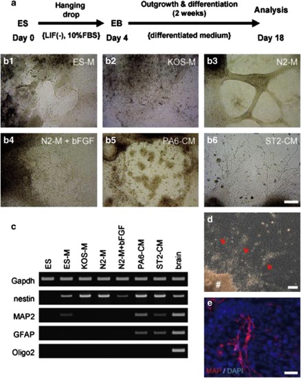
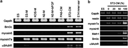
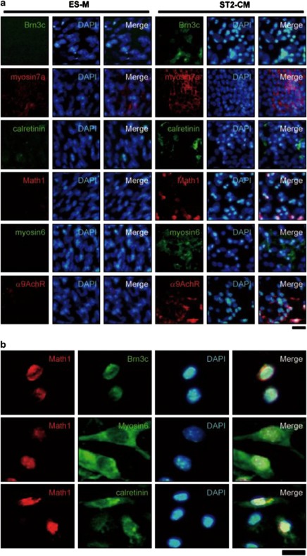
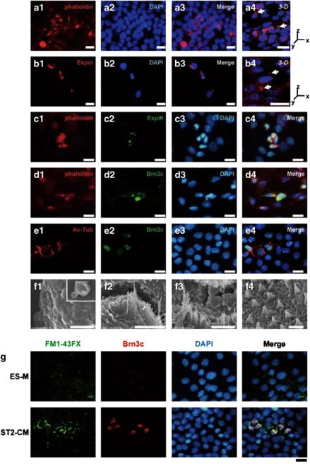
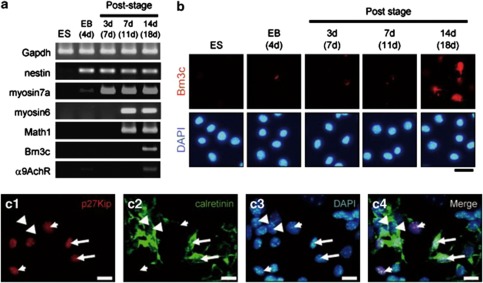
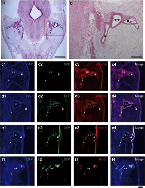
Hearing loss is mainly caused by loss of sensory hair cells (HCs) in the organ of Corti or cochlea. Although embryonic stem (ES) cells are a promising source for cell therapy, little is known about the efficient generation of HC-like cells from ES cells. In the present study, we developed a single-medium culture method for growing embryoid bodies (EBs), in which conditioned medium (CM) from cultures of ST2 stromal cells (ST2-CM) was used for 14-day cultures of 4-day EBs. At the end of the 14-day cultures, up to 20% of the cells in EB outgrowths expressed HC-related markers, including Math1 (also known as Atoh1), myosin6, myosin7a, calretinin, α9AchR and Brn3c (also known as Pou4f3), and also showed formation of stereocilia-like structures. Further, we found that these cells were incorporated into the developing inner ear after transplantation into chick embryos. The present inner ear HC induction method using ST2-CM (HIST2 method) is quite simple and highly efficient to obtain ES-derived HC-like cells with a relatively short cultivation time.


Figures

Figure 1
Figure 1
Directed neural differentiation of ES cells by stromal cells. (a) Schedule of differentiation. Following EB formation, EBs were cultured with various differentiated media (see Table 1) for 2 weeks. (b) Differentiation of EBs was induced using various media, as listed in Table 1. The photographs indicate EB outgrowths after 2 weeks of cultivation: (b-1) ES-M, (b-2) KOS-M, (b-3) N2-M, (b-4) N2-M+bFGF, (b-5) PA6-CM, (b-6) ST2-CM. Scale bar=100 μm. (c) Analysis of expressions neural markers (nestin, MAP2, GFAP, Oligo2) in EB outgrowths after cultivation in various differentiation media for 2 weeks. Samples from adult mouse brain tissues served as a positive control. (d) EB outgrowths cultured with ST2-CM possessed neurite-like structures. The symbol ‘#' and red arrows indicate EB body and neurite-like extension, respectively. Scale bar=100 μm. (e) The neurite-like extensions were immuno-positive for MAP2. Scale bar=20 μm
Figure 2
Figure 2
Gene expressions of HC-related markers in EB outgrowths. (a) Gene expressions of HC-related markers (Brn3c, myosin 6, myosin 7a, and α9AchR) in EB outgrowths cultured with various media were analyzed using a RT-PCR method. Samples from day 14 embryonic (E14) mouse inner ears served as a positive control. (b) Analysis of gene expression of HC-related markers was performed with various concentrations of ST2-CM
Figure 3
Figure 3
Immunocytochemical analysis of specific markers for HCs in EB outgrowths. (a) After 2 weeks of differentiation of EBs in ES-M or ST2-CM, EB outgrowths were analyzed for HC-related markers (Brn3c, myosin7a, calretinin, Math1, myosin6, and α9AchR) using an immunohistochemical method. ST2-CM dramatically induced the emergence of immuno-positive cells for those markers in EB outgrowths. Scale bar=20 μm. (b) Simultaneous expressions of Brn3c, myosin6, and calretinin with Math1 were examined in EB outgrowths cultured with ST2-CM using an immunocytochemical method. Scale bar=10 μm
Figure 4
Figure 4
Stereocilia-like structures formed in HC-like cells. (af) EB outgrowths cultured with ST2-CM for 2 weeks were examined with phalloidin staining and immunostaining for Espin, Brn3c, acetylated tubulin as well as with a SEM. (a) Some cells showed phalloidin-labeled protrusions, highly reminiscent of actin-rich hair bundles (arrow). Scale bars=10 μm. (b) Espin-immuno-positive cells were detected, and the immuno-positivity was found in protrusions at the cell rim (arrow). Scale bars=10 μm. (c) Espin-immuno-positive cells (c-2, green) were simultaneously stained for phalloidin (c-1, red). Scale bars=10 μm. (d) Brn3c-immuno-positive cells (d-2, green) were simultaneously stained for phalloidin (d-1, red). Scale bars=10 μm. (e) Acetylated-tubulin-immuno-positivity (e-1, red) was found in Brn3c-immuno-positive cells (e-2, green). Scale bars=10 μm. (f) Distinct stereocilia-like structures were confirmed by SEM analysis. Shown are undifferentiated ES cells (f-1, low magnification in inset), ES cells differentiated with ST2-CM (f-2 and f-3), and inner ear HCs from the organ of Corti of postnatal mice (day 1) (f-4). Scale bars=3 μm (f-1), 2 μm (f-2), 3 μm (f-3), 5 μm (f-4). (g) The mechanotransduction channel function was examined based on rapid permeation of FM1-43FX dye (green), then immunostaining with Brn3c (red) was performed. Scale bars=20 μm
Figure 5
Figure 5
Recapitulated developmental processes in induction of HC-like cells from ES cells with ST2-CM. (a) Time-course analysis of gene expressions for HC-related markers using RT-PCR method. (b) Time-course analysis of immunocytochemical expression of Brn3c. Scale bar=10 μm. (c) Immunocytochemical analysis of p27kip1 and calretinin in EB outgrowths cultured with ST2-CM for 14 days. Three expression patterns were observed, which were p27kip1-positive and calretinin-negative cells (short arrow), p27kip1-negative and calretinin-positive cells (arrowhead), and p27kip1-positive and calretinin-positive cells (long arrow). Scale bars=10 μm
Figure 6
Figure 6
Transplantation of cells from EB outgrowths containing HC-like cells into chick embryos. (a) Graft cells were transplanted into the otic vesicle of chicken embryos. Three days after injection, normal morphological processes were confirmed by HE staining. Scale bar=1 mm. (b) Enlarged image of area in (a) marked by dotted line. Scale bar=150 μm. (c) No GFP-immuno-positive cells (c-2, green) were found in sections of the inner ear transplanted with undifferentiated ES cells. High phalloidin-positivity was observed in the inner ear (c-3, red). (df) GFP-immuno-positive cells (d-2, e-2, f-2, green) were found to be integrated in the developing inner ear transplanted with cells from EB outgrowths cultured with ST2-CM. Phalloidin-stained cells and GFP-immuno-positive cells were simultaneously observed in the inner ear (d-3, red). (e and f) Most of the integrated cells were immuno-positive for myosin6 (e-3, red) and Brn3c (f-3, red). Scale bar=100 μm. Abbreviations: *cochlear duct; **lateral ampulla; #semicircular canal

References

    1. Carey J, Amin N. Evolutionary changes in the cochlea and labyrinth: Solving the problem of sound transmission to the balance organs of the inner ear. Anat Rec A Discov Mol Cell Evol Biol. 2006;288:482–489. - PubMed
    1. Raphael Y. Cochlear pathology, sensory cell death and regeneration. Br Med Bull. 2002;63:25–38. - PubMed
    1. Fekete DM. Cell fate specification in the inner ear. Curr Opin Neurobiol. 1996;6:533–541. - PubMed
    1. Holley MC. Application of new biological approaches to stimulate sensory repair and protection. Br Med Bull. 2002;63:157–169. - PubMed
    1. Hu Z, Wei D, Johansson CB, Holmström N, Duan M, Frisén J, et al. Survival and neural differentiation of adult neural stem cells transplanted into the mature inner ear. Exp Cell Res. 2005;302:40–47. - PubMed

Publication types

Substances