

Cellular mechanisms of γ-secretase substrate selection, processing and toxicity
- PMID: 22622135
- PMCID: PMC3404154
- DOI: 10.1016/j.pneurobio.2012.05.006
Cellular mechanisms of γ-secretase substrate selection, processing and toxicity
Abstract
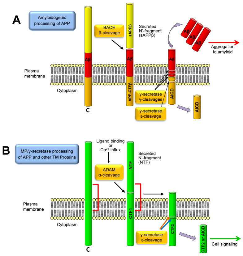
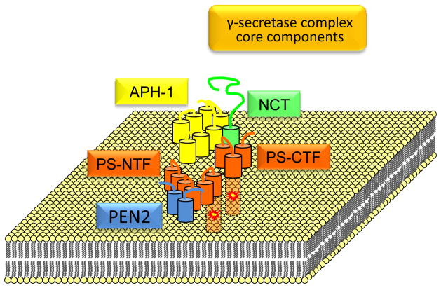
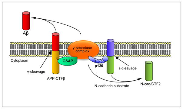
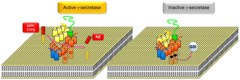
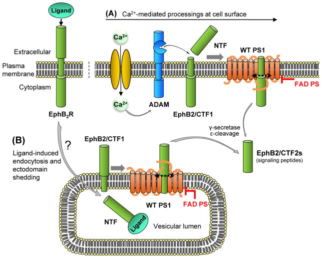
Presenilins (PSs) are catalytic components of the γ-secretase proteolytic complexes that produce Aβ and cell signaling peptides. γ-Secretase substrates are mostly membrane-bound peptides derived following proteolytic cleavage of the extracellular domain of type I transmembrane proteins. Recent work reveals that γ-secretase substrate processing is regulated by proteins termed γ-secretase substrate recruiting factors (γSSRFs) that bridge substrates to γ-secretase complexes. These factors constitute novel targets for pharmacological control of specific γ-secretase products, such as Aβ and signaling peptides. PS familial Alzheimer's disease (FAD) mutants cause a loss of γ-secretase cleavage function at epsilon sites of substrates thus inhibiting production of cell signaling peptides while promoting accumulation of uncleaved toxic substrates. Importantly, γ-secretase inhibitors may cause toxicity in vivo by similar mechanisms. Here we review novel mechanisms that control γ-secretase substrate selection and cleavage and examine their relevance to AD.
Copyright © 2012 Elsevier Ltd. All rights reserved.
Figures
Similar articles
-
Allelic interference: a mechanism for trans-dominant transmission of loss of function in the neurodegeneration of familial Alzheimer's disease.Neurodegener Dis. 2014;13(2-3):126-30. doi: 10.1159/000354241. Epub 2013 Sep 24. Neurodegener Dis. 2014. PMID: 24081144 Free PMC article. Review.
-
Cell signaling abnormalities may drive neurodegeneration in familial Alzheimer disease.Neurochem Res. 2014;39(3):570-5. doi: 10.1007/s11064-013-1003-6. Epub 2013 Feb 23. Neurochem Res. 2014. PMID: 23436150 Free PMC article. Review.
-
Physiological functions of presenilins; beyond γ-secretase.Curr Pharm Biotechnol. 2014;15(11):1019-25. doi: 10.2174/1389201015666141122204139. Curr Pharm Biotechnol. 2014. PMID: 25420727 Review.
-
Presenilin, γ-Secretase, and the Search for Pathogenic Triggers of Alzheimer's Disease.Biochemistry. 2025 Apr 15;64(8):1662-1672. doi: 10.1021/acs.biochem.4c00830. Epub 2025 Feb 25. Biochemistry. 2025. PMID: 39996369 Review.
-
Beyond γ-secretase activity: The multifunctional nature of presenilins in cell signalling pathways.Cell Signal. 2016 Jan;28(1):1-11. doi: 10.1016/j.cellsig.2015.10.006. Epub 2015 Oct 21. Cell Signal. 2016. PMID: 26498858 Review.
Cited by
-
Evidence of a novel mechanism for partial γ-secretase inhibition induced paradoxical increase in secreted amyloid β protein.PLoS One. 2014 Mar 21;9(3):e91531. doi: 10.1371/journal.pone.0091531. eCollection 2014. PLoS One. 2014. PMID: 24658363 Free PMC article.
-
Presenilin-1/γ-secretase controls glutamate release, tyrosine phosphorylation, and surface expression of N-methyl-D-aspartate receptor (NMDAR) subunit GluN2B.J Biol Chem. 2013 Oct 18;288(42):30495-30501. doi: 10.1074/jbc.M113.499004. Epub 2013 Sep 11. J Biol Chem. 2013. PMID: 24025330 Free PMC article.
-
The very many faces of presenilins and the γ-secretase complex.Protoplasma. 2013 Oct;250(5):997-1011. doi: 10.1007/s00709-013-0494-y. Epub 2013 Mar 16. Protoplasma. 2013. PMID: 23504135 Free PMC article. Review.
-
Allelic interference: a mechanism for trans-dominant transmission of loss of function in the neurodegeneration of familial Alzheimer's disease.Neurodegener Dis. 2014;13(2-3):126-30. doi: 10.1159/000354241. Epub 2013 Sep 24. Neurodegener Dis. 2014. PMID: 24081144 Free PMC article. Review.
-
APLP1 as a cerebrospinal fluid biomarker for γ-secretase modulator treatment.Alzheimers Res Ther. 2015 Dec 22;7(1):77. doi: 10.1186/s13195-015-0160-z. Alzheimers Res Ther. 2015. PMID: 26689589 Free PMC article.
References
-
- Andersen OM, Reiche J, Schmidt V, Gotthardt M, Spoelgen R, Behlke J, von Arnim CA, Breiderhoff T, Jansen P, Wu X, Bales KR, Cappai R, Masters CL, Gliemann J, Mufson EJ, Hyman BT, Paul SM, Nykjaer A, Willnow TE. Neuronal sorting protein-related receptor sorLA/LR11 regulates processing of the amyloid precursor protein. Proc Natl Acad Sci U S A. 2005;102:13461–13466. - PMC - PubMed
-
- Anderson JP, Esch FS, Keim PS, Sambamurti K, Lieberburg I, Robakis NK. Exact cleavage site of Alzheimer amyloid precursor in neuronal PC-12 cells. Neurosci Lett. 1991;128:126–128. - PubMed
Publication types
MeSH terms
Substances
Grants and funding
LinkOut - more resources
Full Text Sources
Medical
