. 2012 Sep;13(6):481-8.
doi: 10.1038/gene.2012.22. Epub 2012 May 24.

Lineage-specific adjacent IFNG and IL26 genes share a common distal enhancer element

Affiliations

Lineage-specific adjacent IFNG and IL26 genes share a common distal enhancer element

P L Collins et al. Genes Immun. 2012 Sep.

Abstract

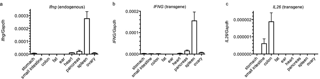
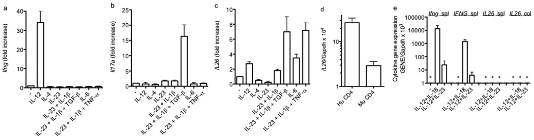
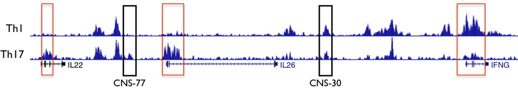
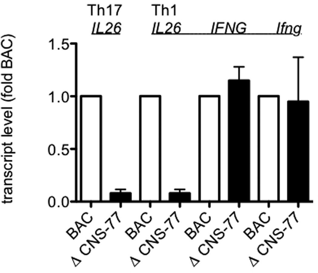
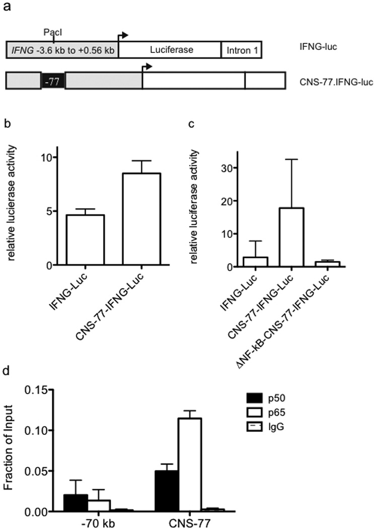
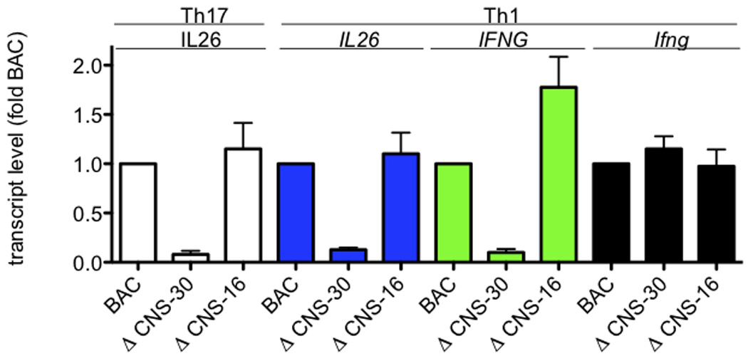
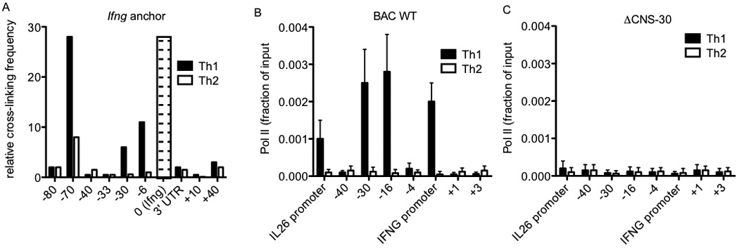
Certain groups of physically linked genes remain linked over long periods of evolutionary time. The general view is that such evolutionary conservation confers 'fitness' to the species. Why gene order confers 'fitness' to the species is incompletely understood. For example, linkage of IL26 and IFNG is preserved over evolutionary time yet Th17 lineages express IL26 and Th1 lineages express IFNG. We considered the hypothesis that distal enhancer elements may be shared between adjacent genes, which would require linkage be maintained in evolution. We test this hypothesis using a bacterial artificial chromosome transgenic model with deletions of specific conserved non-coding sequences. We identify one enhancer element uniquely required for IL26 expression but not for IFNG expression. We identify a second enhancer element positioned between IL26 and IFNG required for both IL26 and IFNG expression. One function of this enhancer is to facilitate recruitment of RNA polymerase II to promoters of both genes. Thus, sharing of distal enhancers between adjacent genes may contribute to evolutionary preservation of gene order.


Conflict of interest statement

Conflict of interest

The authors declare that they have no conflict of interest.

Figures

Figure 1
Figure 1
Basal IFNG and IL26 expression in organs from BAC transgenic mice. Single cell suspensions were prepared from the indicated organs by mechanical disruption or collagenase digestion. Synthesis of cDNA was from purified total RNA. Transcript levels were determined using Taqman assays. Transcript levels of (a) Ifng, (b) IFNG, and (c) IL26 are expressed relative to Gapdh. Error bars are S.D.
Figure 2
Figure 2
Polarizing cytokine requirements for expression of Ifng, Il17a, and IL26 by effector CD4 T cells. CD4 T cells were stimulated with anti-CD3 and anti-CD28 in the presence of the indicated cytokines. After 5 days, cultures were harvested and restimulated with PMA and ionomycin for 6 hrs. Cultures were harvested and transcript levels of (a) Ifng, (b) Il17a, and (c) IL26 were determined as described in Figure 1. Results are expressed relative to cultures stimulated with anti-CD3 and anti-CD28 in the absence of additional cytokines. (d) Human CD4+ Th1 cells (Hu CD4) or murine BAC-transgenic CD4+ Th1 cells (Mu CD4) were stimulated with PMA/Ionomycin. Cultures were harvested and IL26 transcript levels determined as in Figure 1. (e) Murine BAC-transgenic splenic or colonic NK cells (DX5+) were stimulated for 48 hr. Transcript levels of Ifng, IFNG, and IL26 were determined as described in Figure 1. Results are expressed relative to Gapdh transcript levels. Error bars are S.D. * = not detected.
Figure 3
Figure 3
H3K27 acetylation across the IL22-IL26-IFNG gene region in human effector Th1 and Th17 cells. H3K7Ac Chip-Seq data from the ROADMAP ENCODE project were obtained from the U.C.S.C. genome browser. Rectangles in red identify promoter regions. Black rectangles identify two CNS, CNS-77 and CNS-30.
Figure 4
Figure 4
CNS-77 is required for IL26 expression but not IFNG expression. WT and ΔCNS-77 CD4 T cells were stimulated with anti-CD3 and anti-CD28 under Th1 and Th17 polarizing conditions. After 5 days, cultures were harvested and restimulated with PMA and ionomycin for 6 hrs. Transcript levels of IL26, IFNG, and Ifng were determined as described in figure 1. Results are expressed relative to WT BAC levels. Error bars are S.D.
Figure 5
Figure 5
Transcriptional enhancer activity of CNS-77. (a) Schematic of the IFNG-luciferase reporter construct with and without CNS-77 inserted into the Pac1 cloning site. (b) Purified CD4+ T cells were transfected with IFNG-luciferase reporter constructs. After 24 hrs, transfected cells were stimulated with PMA and ionomycin for an additional 24 hrs. Luciferase activity in cell lysates was determined and results are expressed as relative luciferase activity. (c) Purified Jurkat T cells were nucleofected with the indicated reporter constructs and cultured and stimulated as in (b). Luciferase activity in cell lysates was determined and results are expressed as relative luciferase activity. (d) Peripheral human CD4+ T cells were stimulated with anti-CD3 and anti-CD28 for 4 hours and NF-κB binding to CNS-77 was determined by chromatin immunoprecipitation assay. Error bars are S.D.
Figure 6
Figure 6
Both IL26 and IFNG share requirements for CNS-30 for efficient expression. Transgenic IFNG-BAC WT, ΔCNS-30, and ΔCNS-16 CD4 T cells were stimulated with anti-CD3 and anti-CD28 under Th1 or Th17 polarizing conditions. After 5 days, cultures were harvested and restimulated with PMA and ionomycin for 6 hrs. Cultures were harvested and transcript levels of IL26, IFNG, and Ifng were determined as described in Figure 1. Results are expressed relative to WT BAC levels. Error bars are S.D.
Figure 7
Figure 7
RNA polymerase II occupancy across the IL26-IFNG genomic region is dependent upon CNS-30. (a) Average crosslinking frequency between Ifng (indicated by the open bar with dashes) and the indicated genomic positions in nuclei from effector Th1 and Th2 cells. (b & c) RNA pol II recruitment to the indicated genomic positions in (b) BAC WT and (c) ΔCNS-30 transgenic Th1 and Th2 cells determined by ChIP assay. Results are expressed as fraction of input. Error bars are S.D.

References

    1. Boniface K, Blumenschein WM, Brovont-Porth K, McGeachy MJ, Basham B, Desai B, et al. Human Th17 cells comprise heterogeneous subsets including IFN-gamma-producing cells with distinct properties from the Th1 lineage. J Immunol. 2010;185(1):679–687. - PubMed
    1. Cella M, Fuchs A, Vermi W, Facchetti F, Otero K, Lennerz JK, et al. A human natural killer cell subset provides an innate source of IL-22 for mucosal immunity. Nature. 2009;457(7230):722–725. - PMC - PubMed
    1. Chang SH, Dong C. Signaling of interleukin-17 family cytokines in immunity and inflammation. Cell Signal. 2011;23(7):1069–1075. - PMC - PubMed
    1. Commins S, Steinke JW, Borish L. The extended IL-10 superfamily: IL-10, IL-19, IL-20, IL-22, IL-24, IL-26, IL-28, and IL-29. J Allergy Clin Immunol. 2008;121(5):1108–1111. - PubMed
    1. Korn T, Bettelli E, Oukka M, Kuchroo VK. IL-17 and Th17 Cells. Annu Rev Immunol. 2009;27:485–517. - PubMed

Publication types

MeSH terms