. 2012 May 16;485(7399):459-64.
doi: 10.1038/nature11088.

Peroxiredoxins are conserved markers of circadian rhythms

Affiliations

Peroxiredoxins are conserved markers of circadian rhythms

Rachel S Edgar et al. Nature. .

Erratum in

  • Nature. 2012 Sep 27;489(7417):590

Abstract

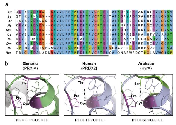
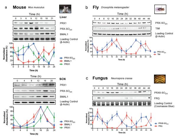
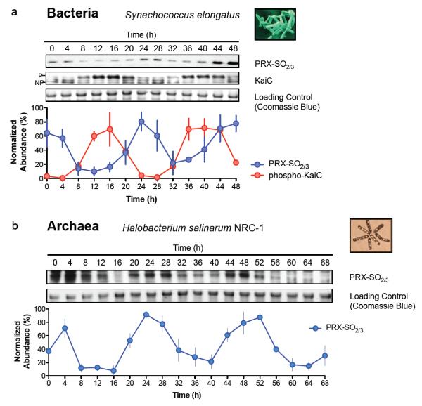
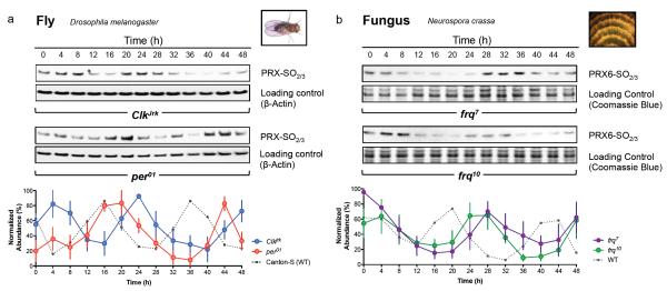
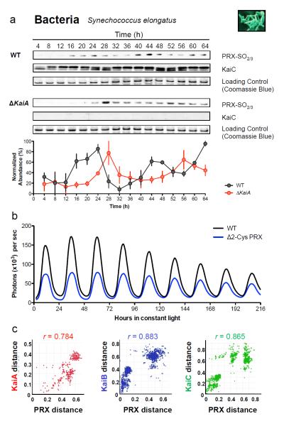
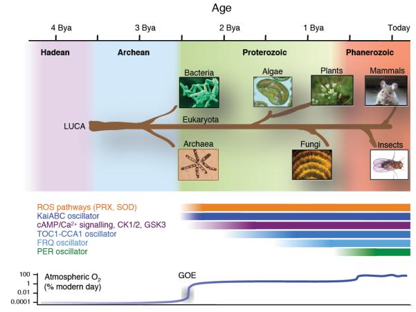
Cellular life emerged ∼3.7 billion years ago. With scant exception, terrestrial organisms have evolved under predictable daily cycles owing to the Earth's rotation. The advantage conferred on organisms that anticipate such environmental cycles has driven the evolution of endogenous circadian rhythms that tune internal physiology to external conditions. The molecular phylogeny of mechanisms driving these rhythms has been difficult to dissect because identified clock genes and proteins are not conserved across the domains of life: Bacteria, Archaea and Eukaryota. Here we show that oxidation-reduction cycles of peroxiredoxin proteins constitute a universal marker for circadian rhythms in all domains of life, by characterizing their oscillations in a variety of model organisms. Furthermore, we explore the interconnectivity between these metabolic cycles and transcription-translation feedback loops of the clockwork in each system. Our results suggest an intimate co-evolution of cellular timekeeping with redox homeostatic mechanisms after the Great Oxidation Event ∼2.5 billion years ago.


Figures

Figure 1
Figure 1. The peroxiredoxin active site is highly conserved in all domains of life
a, Multiple sequence alignment showing peroxiredoxin amino acid sequences. The highly conserved active site is underlined. Representatives shown from Eukaryota (Ot=Ostreococcustauri; At=Arabidopsis thaliana; Hs=Homo sapiens; Mm=Mus musculus; Ce=Caenorhabditis elegans; Sc=Saccharomyces cerevisiae; Dm=Drosophila melanogaster; Nc=Neurospora crassa), Bacteria (Se=Synechococcus elongatus PCC7942) and Archaea (Has=Halobacterium salinarum NRC-1). b, Critical residues in the active site of 2-Cys peroxiredoxins (in bold) are conserved in all organisms. Structures were derived from human PRX-V (PDB code: 1HD2) and human PRDX2 (PDB code: 1QMV), and modified with PyMOL to show the predicted structure for archaeal peroxiredoxin (HyrA, Genbank Accession: NP_280562.1).
Figure 2
Figure 2. Peroxiredoxin oxidation cycles are conserved in eukaryotic models of the circadian clock
Representative immunoblots probed for oxidised/hyperoxidised 2-Cys peroxiredoxin (PRX-SO2/3 or PRX6-SO2/3) are shown for a, mouse (Mus musculus), b, fly (Drosophila melanogaster), and c, fungus (Neurospora crassa). For each model system, the organism was sampled under free-running conditions. Loading controls show either β-actin immunoblots or Coomassie Blue stained gels loaded with identical samples used for immunoblotting. Immunoblot quantification by densitometry is shown below each panel (mean ± SEM) for n=3 biological replicates. See Supplementary Fig. S2 for plant rhythms, and Supplementary Table S9 for cycle period estimates (by harmonic regression) and detailed statistics (by ANOVA).
Figure 3
Figure 3. Peroxiredoxin oxidation cycles are conserved in prokaryotic models of the circadian clock
Representative immunoblots probed for oxidised/hyperoxidised 2-Cys peroxiredoxin (PRX-SO2/3) are shown for a, bacteria (Synechococcus elongatus PCC7942), and b, archaea (Halobacterium salinarum NRC-1). Prior to sampling under free-running conditions (constant light, LL), cyanobacteria were synchronised with a 12 hr dark pulse whereas archaea were stably entrained to 12 hr light: 12 hr dark cycles. Loading controls show Coomassie Blue stained gels loaded with identical samples used for immunoblotting. Immunoblot quantification by densitometry is shown below each panel (mean ± SEM) for n=3 biological replicates. See Supplementary Table S9 for cycle period estimates and detailed statistics. P=phosphorylated KaiC; NP=non-phosphorylated KaiC.
Figure 4
Figure 4. Peroxiredoxin oxidation cycles in circadian clock mutants
Representative immunoblots probed for oxidised/hyperoxidised peroxiredoxin (PRX-SO2/3 or PRX6-SO2/3) are shown for a, fly (Drosophila melanogaster) and b, fungus (Neurospora crassa). For each model system, organisms were sampled under free-running conditions (constant darkness, DD). Loading controls show either β-actin immunoblots or Coomassie Blue stained gels loaded with identical samples used for immunoblotting. Immunoblot quantification by densitometry is shown below each panel (mean ± SEM) for n=3 biological replicates. See Supplementary Table S9 for cycle period estimates (by harmonic regression) and detailed statistics (by ANOVA), as well as Supplementary Fig. S10 for TIM and FRQ immunoblots for fly and fungus respectively.
Figure 5
Figure 5. Relationships between peroxiredoxins and the cyanobacterial Kai-based oscillator
a, Representative immunoblots for oxidised/hyperoxidised 2-Cys peroxiredoxin (PRX-SO2/3) are shown for wild-type (WT; strain AMC149) and KaiA deletion mutant (ΔKaiA; strain AMC702) cyanobacteria. Immunoblot quantification by densitometry is shown below each panel (mean ± SEM), n=3 biological replicates. See Supplementary Table S9 for detailed analysis. b, Bioluminescence traces for cyanobacteria cultures of wild-type (WT) or 2-Cys peroxiredoxin knockout strains (Δ2-Cys PRX), as reported by psbAIp::luxAB. c, Co-evolution of cyanobacteria oscillator components and peroxiredoxin proteins. Interspecies plots correlate inter-protein distances between Kai proteins and 2-Cys peroxiredoxin,. r=correlation coefficient (P< 1×10⁻⁶). See Supplementary Table S8 and Supplementary Fig. S4-6 for details.
Figure 6
Figure 6. Phylogenetic origins of circadian oscillatory systems
A timeline is shown at the top of the schematic (billion years ago, Bya), with the geological era illustrated with a coloured background. A schematic phylogenetic tree shows the origins of each organism studied, stemming from the last universal common ancestor (LUCA). The putative epoch over which each oscillator system has existed is illustrated by the labelled bars. ROS, reactive oxygen species; PRX, peroxiredoxin; SOD, superoxide dismutase; CK1/2, casein kinase 1 or 2; GSK3, glycogen synthase kinase 3.

References

    1. Dunlap JC. Molecular bases for circadian clocks. Cell. 1999;96:271–290. - PubMed
    1. Woelfle MA, Ouyang Y, Phanvijhitsiri K, Johnson CH. The adaptive value of circadian clocks; an experimental assessment in cyanobacteria. Curr Biol. 2004;14:1481–6. - PubMed
    1. Dodd AN, et al. Plant circadian clocks increase photosynthesis, growth, survival, and competitive advantage. Science. 2005;309:630–3. - PubMed
    1. Barger LK, Lockley SW, Rajaratnam SM, Landrigan CP. Neurobehavioral, health, and safety consequences associated with shift work in safety-sensitive professions. Curr Neurol Neurosci Rep. 2009;9:155–64. - PubMed
    1. Wijnen H, Young MW. Interplay of circadian clocks and metabolic rhythms. Annu Rev Genet. 2006;40:409–48. - PubMed

Publication types

MeSH terms