. 2012 May 6;485(7399):507-11.
doi: 10.1038/nature11058.

Sustained translational repression by eIF2α-P mediates prion neurodegeneration

Affiliations

Sustained translational repression by eIF2α-P mediates prion neurodegeneration

Julie A Moreno et al. Nature. .

Erratum in

  • Nature.2014 Jul 17;511(7509):370

Abstract

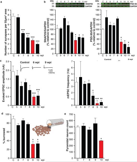
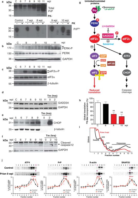
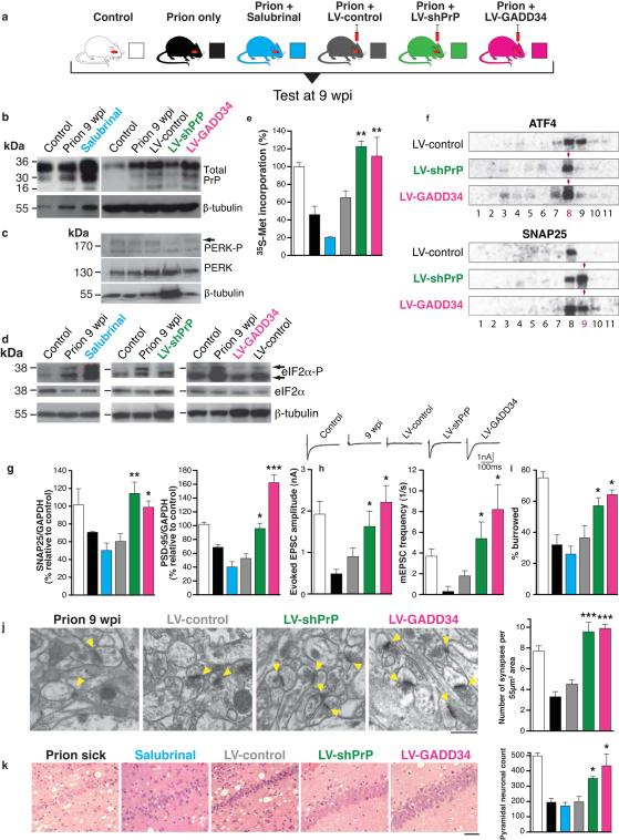
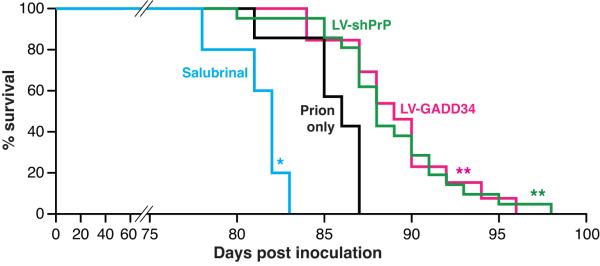
The mechanisms leading to neuronal death in neurodegenerative disease are poorly understood. Many of these disorders, including Alzheimer's, Parkinson's and prion diseases, are associated with the accumulation of misfolded disease-specific proteins. The unfolded protein response is a protective cellular mechanism triggered by rising levels of misfolded proteins. One arm of this pathway results in the transient shutdown of protein translation, through phosphorylation of the α-subunit of eukaryotic translation initiation factor, eIF2. Activation of the unfolded protein response and/or increased eIF2α-P levels are seen in patients with Alzheimer's, Parkinson's and prion diseases, but how this links to neurodegeneration is unknown. Here we show that accumulation of prion protein during prion replication causes persistent translational repression of global protein synthesis by eIF2α-P, associated with synaptic failure and neuronal loss in prion-diseased mice. Further, we show that promoting translational recovery in hippocampi of prion-infected mice is neuroprotective. Overexpression of GADD34, a specific eIF2α-P phosphatase, as well as reduction of levels of prion protein by lentivirally mediated RNA interference, reduced eIF2α-P levels. As a result, both approaches restored vital translation rates during prion disease, rescuing synaptic deficits and neuronal loss, thereby significantly increasing survival. In contrast, salubrinal, an inhibitor of eIF2α-P dephosphorylation, increased eIF2α-P levels, exacerbating neurotoxicity and significantly reducing survival in prion-diseased mice. Given the prevalence of protein misfolding and activation of the unfolded protein response in several neurodegenerative diseases, our results suggest that manipulation of common pathways such as translational control, rather than disease-specific approaches, may lead to new therapies preventing synaptic failure and neuronal loss across the spectrum of these disorders.


Figures

Fig. 1
Fig. 1. Sudden decline of synaptic proteins is the key event leading to synaptic transmission failure and neuronal death in prion-diseased mice
a, Synapse number in the stratum radiatum of the hippocampal CA1 region (imaged by electron microscopy; Supplementary Fig. 1a), declined from 7wpi (n=2 mice; 32 sections per timepoint; p < 0.0001***). b, Levels of pre-synaptic (SNAP-25) and post-synaptic (PSD-95) proteins measured relative to GAPDH declined abruptly at 9wpi. Representative western blots are shown. (n=3 mice per timepoint, p = 0.05*, Student’s t test; 2 tails). c, Whole-cell recordings from CA1 neurons showed significant reduction at 9wpi in amplitude of both evoked excitatory post synaptic currents (EPSC) and frequency of spontaneous miniature mEPSCs. Representative raw traces of evoked EPSCs are shown (left panel, inset). Right panel inset shows amplitude (top) and decay histograms (bottom) showing fewer events per recorded neuron from prion-infected mice, with unchanged mean amplitudes and decay kinetics. (n = 2 mice, 4-8 cells per timepoint. p <0.05* ; p <0.005**, p <0.0001***). d, Burrowing behaviour declines abruptly at 9wpi (n=12 mice; p < 0.001**, Student’s t test; 2 tails) and e, number of CA1 pyramidal neurons is reduced by ~50% at 10wpi (n = 3 mice, 3 sections per mouse; p = 0.04*). All data show mean ± s.e.m. One-way ANOVA with Tukey’s post-test was used unless otherwise stated. Control mice were injected with normal brain homogenate (NBH) and examined at each time point. Data from controls at all time points was averaged, due to lack variability over the time course, to simplify figures. For electrophysiological recordings and EM a single control time point at 10wpi was used.
Fig. 2
Fig. 2. Prion replication induces the unfolded protein response (UPR) and results in eIF2α-P-mediated translational repression
a, Total PrP and PrPSc levels (detected by addition of proteinase K (PK)) increase during prion infection; b, PERK-P and c, eIF2α-P levels rise during disease, d, GADD34 levels do not change but, e, CHOP levels increase throughout disease and f, caspase-12 is cleaved at 10wpi. g, Scheme depicting the translational repression pathway of the UPR showing points for intervention by LV-shPrP, LV-GADD34 and salubrinal. Activation of the pathway results in reduction of global translation at 9wpi, h, determined by 35S-methionine incorporation into hippocampal slices (n = 3 mice, 6 slices; p =0.003**). i, Polysomal profiles from hippocampi show a reduction in active polysomes in fractions 6-11 from prion-infected mice at 9wpi. j, Northern blots on polysomal fractions show increased translation of ATF4 with shift into fraction 10, and decreased translation of SNAP-25 and β-actin (shift into fraction 8) in prion-infected mice at 9wpi. PrP translation is essentially unchanged. Quantitative line plots of Northern blots show percentage of mRNA in each fraction found on the gradients, Tunicamycin (Tm) treated HeLa cells were analysed at 0, 2 and 10 hours as a control for UPR activation. All data show mean ± s.e.m. One-way ANOVA with Tukey’s post-test was used unless otherwise stated. Control mice at 11wpi are shown on western blots, and at 9wpi for Northern blots and 35S met labeling; n = 3 mice for all experiments. For quantification of western blots see Supplementary Fig. 2.
Fig. 3
Fig. 3. Preventing eIF2α-P formation or promoting its dephosphorylation in prion-diseased mice rescues synaptic failure and neuronal loss, while increased eIF2α-P levels exacerbate neurotoxicity
a, Mice were infected with RML prions and treated with salubrinal (blue) or stereotaxically injected with lentiviruses expressing anti-PrP shRNA (LV-shPrP; green) or GADD34 (LV-GADD34; pink) or no insert (LV-control; grey) into both hippocampi. Control groups received no virus (prion only; black) or normal brain homogenate (NBH; control; white). Mice were tested at 9wpi. b, LV-shPrP reduced total PrP and prevented UPR induction, reducing levels of PERK-P c and eIF2α-P, d. LV-GADD34 reduced eIF2α-P despite PERK-P induction, and salubrinal increased eIF2α-P. e, Both LV-GADD34 and LV-shPrP prevented reduction in global translation at 9wpi, but salubrinal reduced translation rates even further. f, LV-GADD34 and LV-shPrP reversed prion-induced eIF2α-P-mediated translational changes of specific mRNAs in polysomal fractions shown on Northern blots. g, Synaptic protein levels; h, synaptic transmission, i, burrowing behaviour, j, and synapse number were protected by GADD34 treatment and PrP knockdown. Salubrinal exacerbated protein loss. Representative EM images (arrowheads denote individual synapses) and quantification are shown (n = 2 mice, 32 slices per mouse for each analysis). k, LV-GADD34 and LV-shPrP resulted in extensive neuroprotection of hippocampal CA1 pyramidal neurons and spongiosis (left hand panels, haematoxylin and eosin stained sections) and chart, right, when the animals were dying of scrapie at nearly 14wpi. Salubrinal accelerated neurodegeneration with extensive neuronal loss seen at 9wpi, earlier than in prion sick animals at 12wpi. Scale bar: 50μm and 2μm for EM images. All data in bar charts show mean ± s.e.m. One-way ANOVA with Tukey’s post test was used for multiple comparisons; p <0.05*; p <0.005**, p <0.0001***. For all experiments n=3 mice, unless otherwise stated. All controls are at 9wpi. For quantification of Northern and western blots see Supplementary Figs. 4 and 9.
Fig. 4
Fig. 4. Reducing eIF2α-P levels in prion-diseased mice significantly increases survival
Kaplan-Meier survival plots for prion-infected mice treated with salubrinal (n=10), prions alone (n=7), hippocampal injections of LV-GADD34 (n=13) or LV-shPrP (n=14). Focal treatment with LV-GADD34 and LV-shPrP resulted in significantly increased survival compared to prion-infected mice with no treatment (p = 0.007**; Student’s t test); salubrinal treatment reduced survival (p =0.03*). For plots for LV-control and DMSO controls see Supplementary Fig. 10.

Comment in

References

    1. Hoozemans JJ, et al. Activation of the unfolded protein response in Parkinson’s disease. Biochem Biophys Res Commun. 2007;354:707–711. - PubMed
    1. Hoozemans JJ, et al. The unfolded protein response is activated in pretangle neurons in Alzheimer′s disease hippocampus. 2009. - PMC - PubMed
    1. Hoozemans JJ, et al. The unfolded protein response is activated in Alzheimer′s disease. Acta Neuropathol. 2005;110:165–172. - PubMed
    1. Unterberger U, et al. Endoplasmic reticulum stress features are prominent in Alzheimer disease but not in prion diseases in vivo. J Neuropathol Exp Neurol. 2006;65:348–357. - PubMed
    1. Boyce M, et al. A selective inhibitor of eIF2alpha dephosphorylation protects cells from ER stress. Science. 2005;307:935–939. - PubMed

Publication types

MeSH terms