. 2012 Apr 29;485(7399):517-21.
doi: 10.1038/nature11007.

Glycolytic oligodendrocytes maintain myelin and long-term axonal integrity

Affiliations

Glycolytic oligodendrocytes maintain myelin and long-term axonal integrity

Ursula Fünfschilling et al. Nature. .

Abstract

Oligodendrocytes, the myelin-forming glial cells of the central nervous system, maintain long-term axonal integrity. However, the underlying support mechanisms are not understood. Here we identify a metabolic component of axon-glia interactions by generating conditional Cox10 (protoheme IX farnesyltransferase) mutant mice, in which oligodendrocytes and Schwann cells fail to assemble stable mitochondrial cytochrome c oxidase (COX, also known as mitochondrial complex IV). In the peripheral nervous system, Cox10 conditional mutants exhibit severe neuropathy with dysmyelination, abnormal Remak bundles, muscle atrophy and paralysis. Notably, perturbing mitochondrial respiration did not cause glial cell death. In the adult central nervous system, we found no signs of demyelination, axonal degeneration or secondary inflammation. Unlike cultured oligodendrocytes, which are sensitive to COX inhibitors, post-myelination oligodendrocytes survive well in the absence of COX activity. More importantly, by in vivo magnetic resonance spectroscopy, brain lactate concentrations in mutants were increased compared with controls, but were detectable only in mice exposed to volatile anaesthetics. This indicates that aerobic glycolysis products derived from oligodendrocytes are rapidly metabolized within white matter tracts. Because myelinated axons can use lactate when energy-deprived, our findings suggest a model in which axon-glia metabolic coupling serves a physiological function.


Figures

Figure 1
Figure 1. Genetic targeting of the mitochondrial COX complex in myelinating glial cells
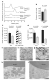
a, Conditional mutagenesis of the floxed Cox10 gene, deleting exon 6 (not drawn to scale). Arrows indicate the location of primers for genotyping (blue/red) and qPCR (red/red), the latter amplifying only the floxed allele. b, Loss of the floxed Cox10 gene in myelinating glial cells of different Cre-expressing mice (adult), confirmed by qPCR of genomic DNA. Highest fraction of myelinating glia are found in sciatic nerve (SN), followed by optic nerve (ON), cervical spinal cord (CSC) and total brain. Mean percentages ± s.d. are shown; n = 3–5. c, Left panel, schematic time line showing that Cox10 genomic recombination in immature oligodendrocytes of newborn Cnp1Cre/+ mice does not interfere with postnatal myelination. Mutant oligodendrocytes myelinate (first 3 weeks), using pre-existing mitochondria that will subsequently decline in respiratory function (marked ‘ageing’). Right panel, for comparison, Cox10 recombination in proliferating Schwann cell precursors perturbs mitochondrial function before myelination, as intact mitochondria are ‘lost’ by dilution with mutant mitochondria. M, month. d, Right panels, myelinated axons and Schwann cells in motor roots of control and mutant animals at P21. The left panel shows a schematic representation. Cox10 mutant Schwann cells (but not axons) lose unstable COX, as demonstrated by reduced immunostaining for the catalytic COX subunit I (brown) in paraffin sections. Schwann cell nuclei (N) are counterstained in pale blue. Upward arrow, COX staining of axonal mitochondria; downward arrow, COX in Schwann cells. Scale bar, 10 μm.
Figure 2
Figure 2. Peripheral neuropathy caused by Cox10 mutant Schwann cells
a, Amplitudes of compound muscle action potentials were recorded in controls and mutant mice at P21 after proximal or distal stimulation of sciatic nerves, but were barely detectable in the mutants. bd, The morphology of sciatic nerves was assessed in semithin sections, and the number of Schwann cell nuclei was counted (b), the cross-sectional area of the nerve with fascicles of axons and Schwann cells was determined (c), and the absolute number of myelinated axons counted (d). This showed a progressive loss of axons at older age, but not of Schwann cells. All data area mean ± s.d. Ctrl, control; mut, mutant. ei, Electron microscopy showed differences between mutant and control sciatic nerves. e, f, In Remak bundles, C-fibre axons (C) are normally sorted by Schwann cell processes (e, arrow) but not in mutants (f).g, Mutant Schwann cells that fail to myelinate survive well. Note numerous unmyelinated medium-calibre axons (A) next to normally myelinated axons (N) and intact Schwann cell nuclei (S). h, i, At higher magnification, myelin appeared morphologically normal, but mutant mitochondria (asterisk) were clearly enlarged. Scale bars, 500 nm (e, f, h, i), 2 μm (g).
Figure 3
Figure 3. Oligodendroglial survival, myelin preservation and white matter integrity inCnp1Cre/+*Cox10flox/flox mice
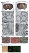
a, b, By Gallyas’ silver impregnation of myelin at 9 months of age, the corpus callosum and other white matter tracts appear normally developed and stable in mutant mice. cf, Electron microscopy of high-pressure frozen optic nerve shows intact myelination of CNS axons (c, d), and healthy oligodendroglia nuclei (e, f). A, axon; N, nucleus; M, mitochondria. Scale bars, 0.5 μm. g, Serial COX and SDH histochemistry. Left, in the normal appearing white matter of 9-month-old Cnp1Cre/+*Cox10flox/flox mutants (corpus callosum), mitochondrial COX activity yields a brown precipitate (axons and astrocytes, for example). By serial COX and SDH histochemistry, only Cox10-mutant COX cells are visibly stained for SDH (blue precipitate, white arrows). Middle, about half (48.2 ± 6.5%) of the OLIG2-positive oligodendrocyte lineage cells in the mutant corpus callosum (red nuclei in overlay), that is, mostly mature oligodendrocytes that express CNP1Cre, are lacking COX activity (blue). Right, there are no COX cells in the corpus callosum of age-matched controls. Scale bar, 20 μm. h, Left, when combining only COX histochemistry with a marker for mature cells (CC1), mutant oligodendrocytes (corpus callosum; 9 months of age) appear white (arrows), intermingled with COX+ neighbouring cells and compartments. Middle, mature oligodendrocytes that are CC1+ (green) are filled with abnormally large numbers of SDH+ mitochondria (red), and appear in orange in the overlay. Note the near absence of SDH signals from neighbouring cells and compartments. Right, in a separate overlay, the SDH+ mitochondria (red) are virtually devoid of COX immunostaining (green) in mutant oligodendrocytes. Scale bar, 10 μm.
Figure 4
Figure 4. Rapid use of lactate shown by proton MRS
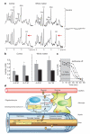
a, Localized proton magnetic resonance spectra of the cortex (left) and corpus callosum (right) fromCnp1Cre/+*Cox10flox/flox mutants and controls (6–7 months of age). Cr, total creatine; Ins, myo-inositol; p.p.m., parts per million; NAA, N-acetylaspartate. Red arrows denote lactate (Lac). b, Lactate levels in the cortex (left) and white matter (right) are increased in mutant mice (grey bars) compared with controls (black bars) under isoflurane anaesthesia. Data are mean ± s.e.m.; n = 6–7 per genotype. Note that under isoflurane anaesthesia the control mice have higher lactate levels in white matter than in grey matter. M, months. c, Increased brain lactate levels drop to undetectable levels in less than 60 min at the end of isoflurane anaesthesia. This suggests that lactate (produced by oligodendrocytes) is rapidly metabolized by other cellular compartments in the white matter tracts of awake mice. d, Hypothetical model of metabolic coupling between oligodendrocytes and myelinated axons. Oligodendrocytes import glucose through GLUT1 (and possibly via astrocytes and gap junctions; CX, connexin) for glycolysis. Pyruvate is metabolised in mitochondria (yellow) for ATP generation (TCA, tricarboxylic acid cycle). With the onset of myelination (‘developmental switch’), glucose also serves the synthesis of fatty acid (FAS) and myelin lipids from acetyl-CoA. In post-myelination oligodendrocytes, glycolysis can yield sufficient ATP to support oligodendrocyte survival. Glycolysis products are used by myelinated axons when energy levels are low,. Lactate (or pyruvate when NADH is oxidized in oligodendroglial mitochondria) can be directly transferred via monocarboxylic acid transporters (MCT1, MCT2), which reside in internodal myelin and the axonal compartment, such that lactate is rapidly cleared in vivo (in c). Note that myelinated axons, largely shielded from the extracellular milieu, are separated by a thin periaxonal space from the oligodendroglial cytoplasm filling the inner loops of myelin (‘cytosolic channel’) and paranodal loops.

References

    1. Griffiths I, et al. Axonal swellings and degeneration in mice lacking the major proteolipid of myelin. Science. 1998;280:1610–1613. - PubMed
    1. Lappe-Siefke C, et al. Disruption of Cnp1 uncouples oligodendroglial functions in axonal support and myelination. Nature Genet. 2003;33:366–374. - PubMed
    1. Kassmann CM, et al. Axonal loss and neuroinflammation caused by peroxisome-deficient oligodendrocytes. Nature Genet. 2007;39:969–976. - PubMed
    1. Nave KA. Myelination and the trophic support of long axons. Nature Rev. Neurosci. 2010;11:275–283. - PubMed
    1. Ziabreva I, et al. Injury and differentiation following inhibition of mitochondrial respiratory chain complex IV in rat oligodendrocytes. Glia. 2010;58:1827–1837. - PMC - PubMed

Publication types

MeSH terms