. 2012 Jul;91(7):683-9.
doi: 10.1177/0022034512449170. Epub 2012 May 23.

Muenke syndrome mutation, FgfR3P²⁴⁴R, causes TMJ defects

Affiliations

Muenke syndrome mutation, FgfR3P²⁴⁴R, causes TMJ defects

T Yasuda et al. J Dent Res. 2012 Jul.

Abstract

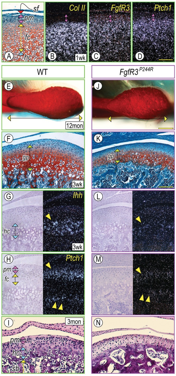
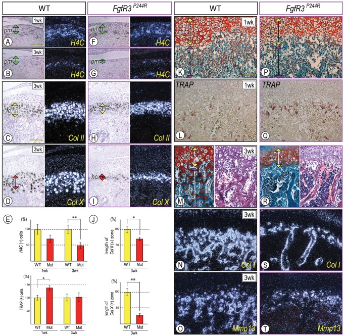
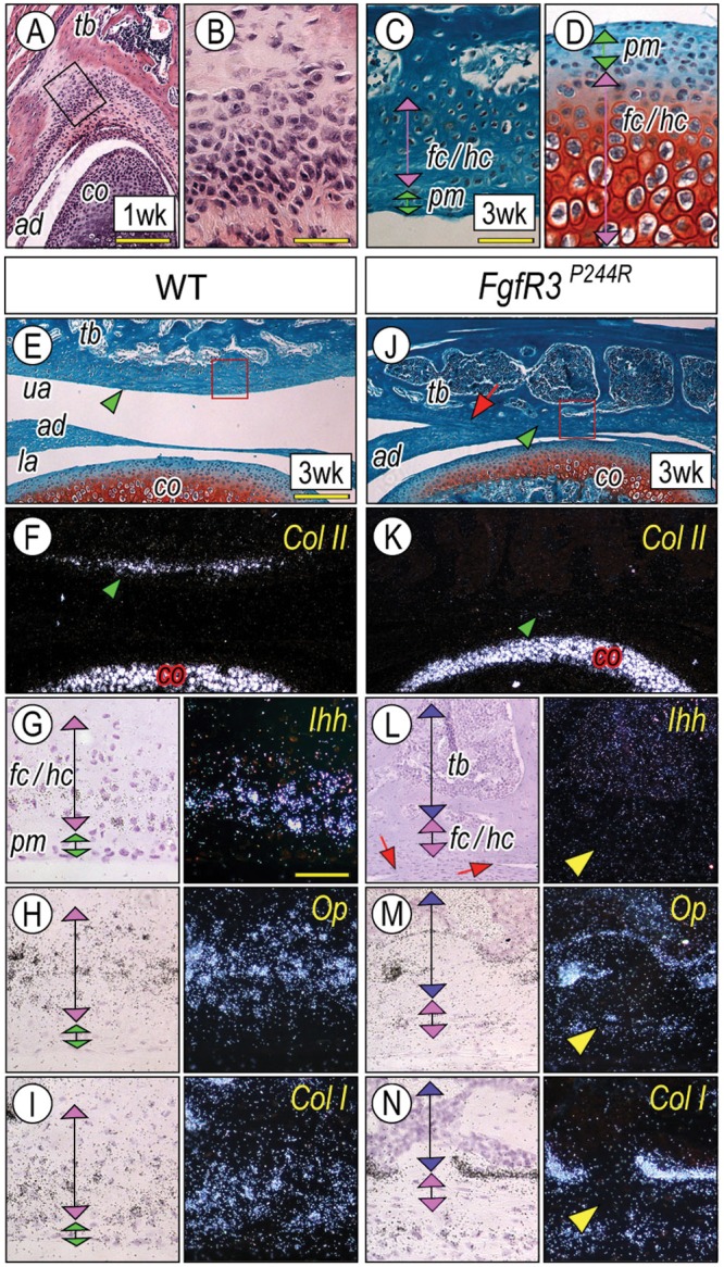
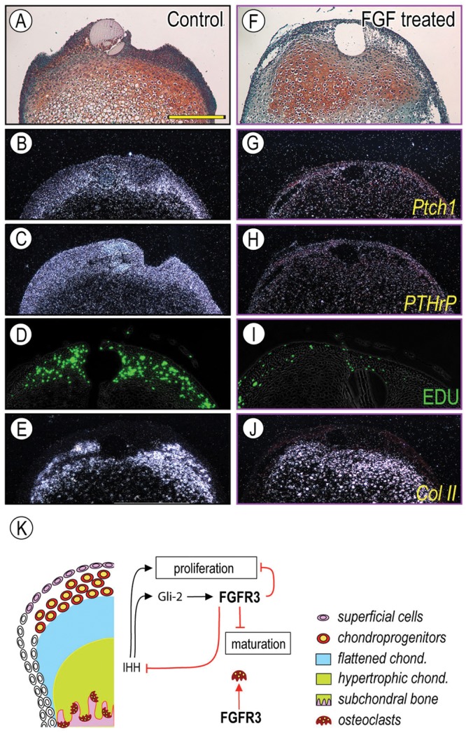
Muenke syndrome is characterized by various craniofacial deformities and is caused by an autosomal-dominant activating mutation in fibroblast growth factor receptor 3 (FGFR3(P250R) ). Here, using mice carrying a corresponding mutation (FgfR3(P244R) ), we determined whether the mutation affects temporomandibular joint (TMJ) development and growth. In situ hybridization showed that FgfR3 was expressed in condylar chondroprogenitors and maturing chondrocytes that also expressed the Indian hedgehog (Ihh) receptor and transcriptional target Patched 1(Ptch1). In FgfR3(P244R) mutants, the condyles displayed reduced levels of Ihh expression, H4C-positive proliferating chondroprogenitors, and collagen type II- and type X-expressing chondrocytes. Primary bone spongiosa formation was also disturbed and was accompanied by increased osteoclastic activity and reduced trabecular bone formation. Treatment of wild-type condylar explants with recombinant FGF2/FGF9 decreased Ptch1 and PTHrP expression in superficial/polymorphic layers and proliferation in chondroprogenitors. We also observed early degenerative changes of condylar articular cartilage, abnormal development of the articular eminence/glenoid fossa in the TMJ, and fusion of the articular disc. Analysis of our data indicates that the activating FgfR3(P244R) mutation disturbs TMJ developmental processes, likely by reducing hedgehog signaling and endochondral ossification. We suggest that a balance between FGF and hedgehog signaling pathways is critical for the integrity of TMJ development and for the maintenance of cellular organization.


Conflict of interest statement

The authors declare no potential conflicts of interest with respect to the authorship and/or publication of this article.

Figures

Figure 1.
Figure 1.
Mandibular condyle development was defective in FgfR3P244R mutant mice. Parasagittal sections from wild-type P7 (A-D), P21 (F-H), 3-month (I) and mutant P21 (K-M), and 3-month (N) condyles were processed for safranin-O/fast green staining (A, F, K), hematoxylin-eosin staining (I, N), and in situ hybridization (B-D, G-H, L-M). Twelve-month-old control (E) and mutant (J) condyles were stained with Alcian blue/alizarin red. In situ hybridization with the isotope-labeled RNA probes for collagen II (Col II) (B), Fibroblast growth factor Receptor 3 (FgfR3) (C), and Patched1 (Ptch1) (D). Note that FgfR3 expression largely overlapped with Ptch1-expressing cells in the polymorphic cell layer (pm) and flattened chondrocyte zone (fc). Superficial view of control (E) and mutant (J) condyles; mutant condyle is hypomorphic, indicated by shortening along the antero-posterior axis (a horizontal double arrow) compared with control (E). Note that P21-mutant condyles (K) exhibited the shorter growth-plate-like structure (gp, a green vertical double arrow) compared with controls (F). Ihh transcripts in the hypertrophic chondrocyte layer (hc) (G, arrowhead) and Ptch1 mRNAs in the polymorphic cell layer (pm, a purple vertical double arrow), the flattened chondrocyte layer (fc, a yellow vertical double arrow) (H, arrowhead), and the developing trabecular bones (H, a double arrowhead). Note the significant decrease of Ihh (L) and Ptch1 (M) transcripts in P21 mutant condyles, and chondroprogenitors and hypertrophic chondrocytes in 3-month mutant condyles (N). Trabecular bone with large marrow cavities underneath articular cartilage was appreciable in mutant condyles (N). sf, superficial layer; pm, polymorphic layer; fc, flattened chondrocyte layer; hc, hypertrophic chondrocyte layer. Scale bars: 120 µm for A-D (in D), 0.5 mm for E and J (in J), and 150 µm for F-I and K-N (in K).
Figure 2.
Figure 2.
Chondroprogentor proliferation, growth plate organization, and endochondral ossification were abnormal in FgfR3P244R mutant condyles. Parasagittal sections from control P7 (A, K, L) and P21 (B-D, M-O) condyles and mutant P7 (F, P, Q) and P21 (G-I, R-T) condyles were processed for safranin O/fast green staining (K, M, P, R), hematoxylin and eosin staining (M, R), in situ hybridization (A-D, F-I, N, O, S, T), and TRAP staining (L, Q). In situ hybridization with the isotope-labeled RNA probes for histone 4C (H4C) (A, B, F, G), and collagen type II (Col II) (C, H) and type X (Col X) (D, I). Note that, in P21 mutants, condyles displayed a significant reduction of H4C-expressing proliferating chondroprogenitors in the polymorphic layer (pm) (G), and the Col II-expressing flattened chondrocyte layer (fc) (H) and Col X-expressing hypertrophic chondrocyte layer (hc) (I) became narrower. For quantification of the proliferating chondroprogenitors (E), distinct condylar sections (approximately 100 cells/polymorphic layer, n = 6) from each of P7/P21 control and mutant mice were chosen, proliferating cells were present as the ratio/total cells, and each control was set as 100%. For quantification of the length of flattened and hypertrophic zones, Col II-expressing and Col X-expressing cells along the longitudinal axis were counted in distinct condylar sections (n = 6) from P21 control and mutant mice. The number of each control was set as 100%. p-values less than 0.05 were considered as statistically significant (*p < 0.05, **p < 0.02). Note that TRAP-positive osteoclasts were increased in the chondro-osseous border in P7 mutant condyles (E, Q; *p < 0.02) without detection of the clear histomorphogical changes of subchondral bone formation (sb) (P) compared with control (K, L, respectively). Note, in P21 mutant condyles, the significant reduction in bony spicules in the subchondral bone (sb) (R), accompanied by decreased Col I/Mmp13 (S, T)-expressing osteoblasts in the trabecular bone. pm, polymorphic layer; fc, flattened chondrocyte layer; hc, hypertrophic chondrocyte layer; sb, subchondral bone. Scale bars: 120 µm for A, B, F, G (in A); 90 µm for C, D, H, I (in C); and 90 µm for K-T (in K).
Figure 3.
Figure 3.
A unique process of chondrogenesis in the articular eminence/glenoid fossa and the abnormal cellular organization in FgfR3P244R-mutant mice. Serial frontal sections from wild-type P7 (A, B) and P21(C) articular eminence/glenoid fossa and P21 mandibular condyles (D) were processed for hematoxylin-eosin (A, B) and safranin O/fast green (C, D) staining. The magnified image (B) from the boxed area in (A) shows that the mesenchymal condensation of the articular eminence/glenoid fossa first emerged in the thickened periosteum of the temporal bone (tb) after the condyle (co) and articular disc (ad) formations had taken place. Note that polymorphic (pm) and differentiating chondrocyte (fc/hc) layers of the articular eminence/glenoid fossa (C) are narrower and hypertrophic chondrocytes are smaller than those in the mandibular condyles (D). Serial sections from control P21 (E-I) and mutant (J-N) TMJ were processed for safranin-O/fast green staining (E, J) and in situ hybridization with indicated probes (F-I, K-N). The magnified image (G-I, L-N) from the boxed area in E and J, respectively, shows the developing articular eminence/glenoid fossa. Note the absence of Col II transcripts (K, arrowhead) in the mutant articular eminence/glenoid fossa. Note also the articular disc fusion to the temporal bone (J, L, arrows). Scale bars: 120 µm in A, 45 µm in B, 60 µm for C, D (in C), 180 µm for E, F, J, K (in E), and 50 µm for G-I and L-N (in G).
Figure 4.
Figure 4.
FGF signaling decreased expression of Ihh downstream targets and proliferation of chondroprogenitors in the polymorphic layer. Control BSA-treated (A-E) and rFGF9-treated (F-J) condyles were processed for in situ hybridization with isotope-labeled RNA probes for Patched1 (Ptch1) (B, G), PTHrP (C, H), and Collagen II (Col II) (E, J), and for proliferation detection (D, I). In control condyles, Ptch1 and PTHrP were expressed in the superficial/polymorphic layer (B, C), and EDU identifies proliferating chondroprogenitors in the polymorphic layers (D). Note that the rFGF9 treatment reduced Ptch1 transcripts in the superficial/polymorphic layers (G, H, respectively) and EDU incorporation (I), consistent with our observation in FgfR3P244R mutant mice and indicating that rFGF9 acted by inhibiting Ihh expression or signaling. Scale bar: 120 µm for A-J (in A). Schematic depicting our current working model for FGF-Ihh signaling interactions in post-natal TMJ is shown in (K). Ihh signaling regulated proliferation of chondroprogenitors (Shibukawa et al., 2007; Ochiai et al., 2010), and Gli2 inhibits chondrocyte hypertrophy via FgfR3 signaling (Purcell et al., 2009). FGFR3 signaling affects osteoclast differentiation/distribution. A similar molecular regulation could apply to the secondary cartilage in the articular eminence/glenoid fossa. ae/gf, articular eminence/glenoid fossa; ad, articular disc.

References

    1. Colvin JS, Bohne BA, Harding GW, McEwen DG, Ornitz DM. (1996). Skeletal overgrowth and deafness in mice lacking fibroblast growth factor receptor 3. Nat Genet 12:390-397 - PubMed
    1. Deng C, Wynshaw-Boris A, Zhou F, Kuo A, Leder P. (1996). Fibroblast growth factor receptor 3 is a negative regulator of bone growth. Cell 84:911-921 - PubMed
    1. Hinton RJ, McNamara JA., Jr (1984). Temporal bone adaptations in response to protrusive function in juvenile and young adult rhesus monkeys (Macaca mulatta). Eur J Orthod 6:155-174 - PubMed
    1. Ibrahimi OA, Zhang F, Eliseenkova AV, Linhardt RJ, Mohammadi M. (2004). Proline to arginine mutations in FGF receptors 1 and 3 result in Pfeiffer and Muenke craniosynostosis syndromes through enhancement of FGF binding affinity. Hum Mol Genet 13:69-78 - PubMed
    1. Kantomaa T, Tuominen M, Pirttiniemi P. (1994). Effect of mechanical forces on chondrocyte maturation and differentiation in the mandibular condyle of the rat. J Dent Res 73:1150-1156 - PubMed

Publication types

MeSH terms

Substances

Supplementary concepts

LinkOut - more resources