

Middle ear adenomas stain for two cell populations and lack myoepithelial cell differentiation
- PMID: 22623086
- PMCID: PMC3422582
- DOI: 10.1007/s12105-012-0365-4
Middle ear adenomas stain for two cell populations and lack myoepithelial cell differentiation
Abstract
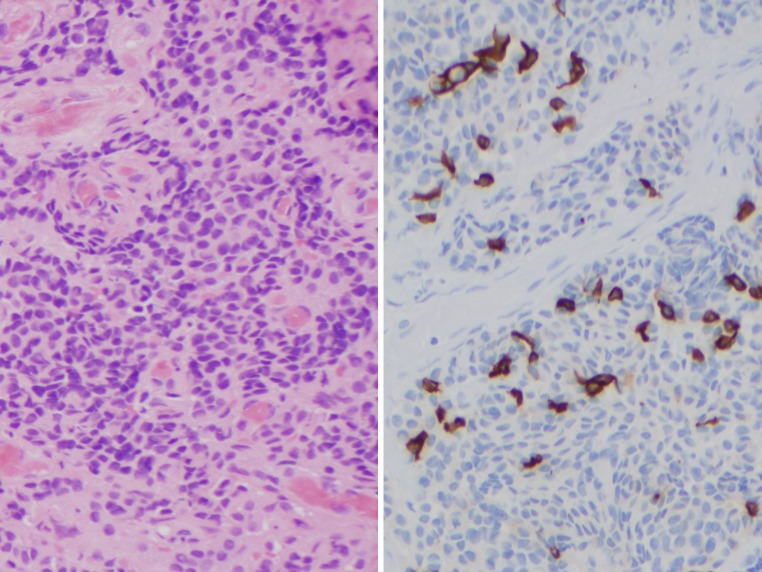
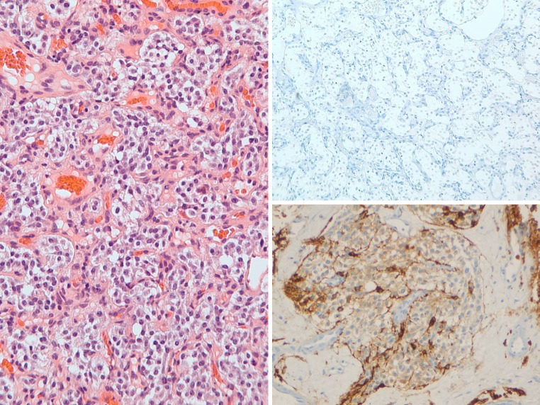
Middle ear adenomas (MEAs) are benign neoplasms along a spectrum with neuroendocrine neoplasms (carcinoid tumors). Immunohistochemical (IHC) staining for myoepithelial markers has not been reported in these tumors. The archives of the Cleveland Clinic, University of Virginia and Armed Forces Institute of Pathology were retrospectively searched for tumors arising within the middle ear with material available for IHC staining. Twelve cases of MEAs, four cases of jugulotympanic paragangliomas (JPGs), 10 cases of ceruminous adenomas (CAs) and four cases of ceruminous adenocarcinomas (CACs) were obtained. IHC staining was performed for smooth muscle actin (SMA), p63, S-100 protein, cytokeratin 5/6 (CK5/6), and cytokeratin 7 (CK7). The MEAs were positive for: CK7 (92 %, luminal), CK5/6 (92 %, abluminal), p63 (83 %, abluminal), and negative for SMA and S-100 protein. The JPGs were negative for CK7, CK5/6, p63 and SMA; S-100 protein highlighted sustentacular cells. The CAs were positive for: CK7 (100 %, luminal), CK5/6 (100 %, abluminal), S-100 protein (80 %, abluminal), p63 (100 %, abluminal), and SMA (90 %, abluminal). CACs demonstrated two patterns, (1) adenoid cystic carcinoma-type: positive for CK7 (100 %, luminal), CK5/6, S-100 protein, p63, and SMA (all 100 %, abluminal); and (2) conventional-type: CK7 (50 % luminal), and no CK5/6, SMA, S-100 protein, or p63 expression. The IHC profile of MEAs suggests that these tumors harbor at least two cell populations, including luminal and basal cells. However, unlike ceruminous adenomas, MEAs lack true myoepithelial differentiation given the absence of S-100 protein and SMA staining in all cases.
Figures


References
-
- Berns S, Pearl G. Middle ear adenoma. Arch Pathol Lab Med. 2006;130:1067–1069. - PubMed
MeSH terms
LinkOut - more resources
Full Text Sources
Research Materials
