. 2012 Jul;7(7):1286-94.
doi: 10.1002/cmdc.201200104. Epub 2012 May 23.

Structural characterization and computer-aided optimization of a small-molecule inhibitor of the Arp2/3 complex, a key regulator of the actin cytoskeleton

Affiliations

Structural characterization and computer-aided optimization of a small-molecule inhibitor of the Arp2/3 complex, a key regulator of the actin cytoskeleton

Andrew W Baggett et al. ChemMedChem. 2012 Jul.

Abstract

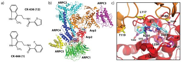
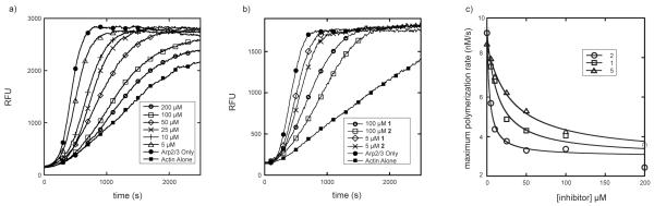
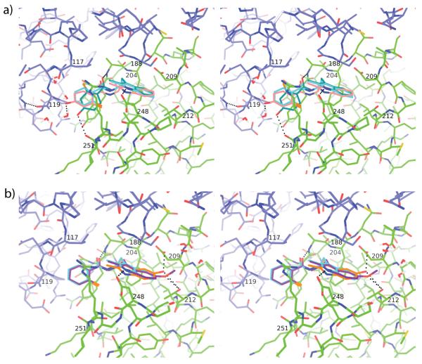
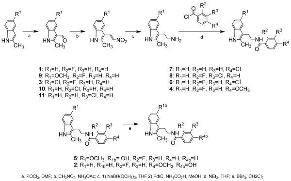
CK-666 (1) is a recently discovered small-molecule inhibitor of the actin-related protein 2/3 (Arp2/3) complex, a key actin cytoskeleton regulator with roles in bacterial pathogenesis and cancer cell motility. Although 1 is commercially available, the crystal structure of Arp2/3 complex with 1 bound has not been reported, making its mechanism of action uncertain. Furthermore, its relatively low potency increases its potential for off-target effects in vivo, complicating interpretation of its influence in cell biological studies and precluding its clinical use. Herein we report the crystal structure of 1 bound to Arp2/3 complex, which reveals that 1 binds between the Arp2 and Arp3 subunits to stabilize the inactive conformation of the complex. Based on the crystal structure, we used computational docking and free-energy perturbation calculations of monosubstituted derivatives of 1 to guide optimization efforts. Biochemical assays of ten newly synthesized compounds led to the identification of compound 2, which exhibits a threefold increase in inhibitory activity in vitro relative to 1. In addition, our computational analyses unveiled a surface groove at the interface of the Arp2 and Arp3 subunits that can be exploited for additional structure-based optimization.


Figures

Figure 1
Figure 1
(A) Previously synthesized Arp2/3 complex inhibitors 1 and 12. Compound 1 was used as a parent scaffold for substitutions indicated in Scheme 1. (B) Crystal structure of 1 bound to Bos taurus Arp2/3 complex. 1 is shown as spheres and binds at the interface of the Arp3 (orange) and Arp2 (red) subunits. The five other subunits in the complex are labeled ARPC1-5. (C). Comparison of 1 (grey carbon atoms) with 12 (turquoise carbon atoms) showing a close up of the binding pocket at the interface of Arp2 and Arp3. Coordinates for 12 for this figure were generated by overlaying Arp2 and Arp3 from 3DXK.pdb onto the structure of 1 and applying the transformation to 12.
Figure 2
Figure 2
Biochemical characterization of 1 and analogues. (A) Time course of the polymerization of 30% pyrene-labeled actin in the presence of 5 nM Arp2/3 complex, 200 nM N-WASP-VCA, and either DMSO for a range of concentrations of 1. A control reaction (actin alone) lacking only Arp2/3 complex shows the intrinsic nucleation rate of actin. (B) Comparison of actin polymerization time courses in the presence of equal concentrations of 1 versus 2. Conditions are identical to panel (A). (C) Plot of maximal polymerization rate versus inhibitor concentration for 1, 2, and 5. Data were fit as described in Methods.
Figure 3
Figure 3
Binding pocket of 1 with overlapping docked analogues 2, 5 and 9. (A) Stereo figure showing a comparison of 1 and the docked and FEP poses of 2. Arp3 (blue carbon atoms) and Arp2 (green carbon atoms) from the crystal structure of Arp2/3 with bound 1 (3UKR.pdb, light blue carbon atoms) are shown in line representation overlaid with the Glide docked pose (teal carbon atoms) and the FEP docked pose (pink) of 2. Selected hydrogen bonds are shown as black dotted lines. (B) Stereo figure showing a comparison of 1 and the FEP generated poses of 5 and 9. Colors are identical to panel A, except 5 is shown with orange carbon atoms and 9 is shown with purple carbon atoms.
Scheme 1
Scheme 1

References

    1. Pollard TD, Cooper JA. Science. 2009;326(5957):1208–1212. - PMC - PubMed
    1. Cooper JA, Buhle EL, Jr, Walker SB, Tsong TY, Pollard TD. Biochemistry. 1983;22(9):2193–2202. - PubMed
    1. Firat-Karalar EN, Welch MD. Curr Opin Cell Biol. 2011;23(1):4–13. - PMC - PubMed
    1. Goley ED, Welch MD. Nat Rev Mol Cell Biol. 2006;7(10):713–726. - PubMed
    1. Sawa M, Suetsugu S, Sugimoto A, Miki H, Yamamoto M, Takenawa T. J Cell Sci. 2003;116(Pt 8):1505–1518. - PubMed
    2. Hudson AM, Cooley L. J Cell Biol. 2002;156(4):677–687. - PMC - PubMed

Publication types

MeSH terms

Substances