. 2012 May;24(5):1746-59.
doi: 10.1105/tpc.112.097998. Epub 2012 May 22.

Screening a cDNA library for protein-protein interactions directly in planta

Affiliations

Screening a cDNA library for protein-protein interactions directly in planta

Lan-Ying Lee et al. Plant Cell. 2012 May.

Abstract

Screening cDNA libraries for genes encoding proteins that interact with a bait protein is usually performed in yeast. However, subcellular compartmentation and protein modification may differ in yeast and plant cells, resulting in misidentification of protein partners. We used bimolecular fluorescence complementation technology to screen a plant cDNA library against a bait protein directly in plants. As proof of concept, we used the N-terminal fragment of yellow fluorescent protein- or nVenus-tagged Agrobacterium tumefaciens VirE2 and VirD2 proteins and the C-terminal extension (CTE) domain of Arabidopsis thaliana telomerase reverse transcriptase as baits to screen an Arabidopsis cDNA library encoding proteins tagged with the C-terminal fragment of yellow fluorescent protein. A library of colonies representing ~2 × 10(5) cDNAs was arrayed in 384-well plates. DNA was isolated from pools of 10 plates, individual plates, and individual rows and columns of the plates. Sequential screening of subsets of cDNAs in Arabidopsis leaf or tobacco (Nicotiana tabacum) Bright Yellow-2 protoplasts identified single cDNA clones encoding proteins that interact with either, or both, of the Agrobacterium bait proteins, or with CTE. T-DNA insertions in the genes represented by some cDNAs revealed five novel Arabidopsis proteins important for Agrobacterium-mediated plant transformation. We also used this cDNA library to confirm VirE2-interacting proteins in orchid (Phalaenopsis amabilis) flowers. Thus, this technology can be applied to several plant species.


Figures

Figure 1.
Figure 1.
Sensitivity of the BiFC Reaction in Arabidopsis Leaf Protoplasts. (A) Schematic representation of the constructions used in BiFC. Expression of VirE2-nVenus/nYFP is under the control of the relatively weak nopaline synthase (nos) promoter, whereas expression of VirE2-cCFP, cYFP-EV, or cYFP-cDNA protein is under the control of the strong CaMV double 35S promoter. Term35S, CaMV 35S poly(A) addition signal. Interaction of VirE2-nVenus/nYFP with VirE2-cCFP or with a cYFP-cDNA–encoded protein may permit the two YFP-derived fragments to fold, generating yellow fluorescence. (B) Interaction of various quantities of a plasmid encoding VirE2-cCFP (diluted with the EV plasmid) and 20 μg of a plasmid encoding VirE2-nVenus. The three plasmids were cotransfected into Arabidopsis leaf protoplasts and visualized after 16 h by confocal microscopy. For each set of images: top left, YFP fluorescence; top right, chlorophyll autofluorescence; bottom left, differential interference contrast image; bottom right, merged yellow fluorescence and chlorophyll autofluorescence images.
Figure 2.
Figure 2.
Reconstruction Reaction to Determine the Sensitivity of VirE2-cDNA Clone Interaction in BiFC. (A) Arabidopsis leaf protoplasts were cotransfected with plasmids encoding mRFP-VirD2NLS (to mark transfected cells), VirE2-nVenus (10 μg; bait), and a total of 10 μg of a combination of a cDNA encoding a HSR201-like protein (At4g15390; the prey) plus cYFP-RPN9 (At5g45620; noninteracting protein) in various ratios (indicated on the abscissa). The cells were imaged by confocal microscopy after 16 h. The graph depicts the percentage of cells that were transfected (mRFP fluorescence) and the ratio of cells showing YFP:mRFP fluorescence. Note that the transfection efficiency was approximately the same in all experiments and that as the ratio of cYFP-HSR201-like protein:HSR201-like+RPN9 increases, a higher percentage of cells display YFP fluorescence. (B) Merged confocal images of representative fields of transfected protoplasts. Ratios as in (A). Red, mRFP fluorescence; yellow, YFP BiFC fluorescence; blue, false-color chlorophyll image. Bars = 50 μm.
Figure 3.
Figure 3.
Subcellular Localization of YFP Fluorescence Generated by Interaction of VirE2-nVenus with Different cYFP-Tagged cDNAs. Arabidopsis leaf protoplasts were transfected with 20 μg each of plasmids encoding VirE2-nVenus and a cYFP-tagged cDNA library. The cells were imaged by confocal microscopy 16 h later. Panels show examples of perinuclear (A) and nuclear (B) BiFC signals. White broken circle indicates the nucleus, which was identified visually from other images in the Z-stack. DIC, differential interference contrast. Bars = 20 μm.
Figure 4.
Figure 4.
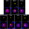
Interaction of nYFP-VirD2 with Various Proteins. Arabidopsis leaf protoplasts were transfected with 10 μg each of plasmids encoding nYFP-VirD2, specific cYFP-tagged cDNA clones, and mCherry. The cells were imaged by confocal microscopy 16 h later. Top panels from each set show yellow YFP fluorescence images. Bottom panels show overlay images of YFP fluorescence (yellow), mCherry fluorescence (red; marks both the cytoplasm and nucleus), and chloroplasts (blue pseudocolor). Individual plasmids used for interaction encode the following: RPN9 (26S proteasome regulatory subunit RPN9 [At5g45620]; negative control for a protein that does not interact with VirD2); IMPa-3 (At4g02150); GTPase (putative GTPase [At3g07050]); MYST-like HAT2 (MYST-like histone acetyltransferase 2 [At5g09740]); HOG1 (S-adenosyl-l-homocysteine hydrolase [At4g13940]); ROC3 (cyclophilin ROC3 [At2g16600]); and RBP 45A (RNA binding protein 45A [At5g54900]). Bars = 10 μm.
Figure 5.
Figure 5.
Mutation of Several VirD2- and VirE2-Interacting Proteins Results in Altered Susceptibility to Agrobacterium-Mediated Root Transformation. Two independent alleles of Arabidopsis At4g15390, encoding a HSR-201–like protein that interacts with VirE2 (A); two independent alleles of At5g09740, encoding a MYST-like histone acetyltransferase 2 that interacts with VirD2 (B); At3g07050, encoding a putative GTPase that interacts with VirD2, two independent alleles of At5g54900, encoding the RNA binding protein 45A that interacts with VirD2, and two independent alleles of At4g13940, hog1-4, and sah1, encoding a S-adenosyl-l-homocysteine hydrolase that interacts with both VirD2 and VirE2 (C). Root segments from wild-type Arabidopsis and various mutants were inoculated with the indicated concentrations of the tumorigenic strain Agrobacterium A208 (for stable transformation) or the disarmed strain At849 (for transient β-glucuronidase expression) as described (Tenea et al., 2009). The percentage (of control) of root segments generating crown gall tumors ([A] and the left side of [B] and [C]) was scored after 1 month of growth on MS medium lacking phytohormones for tumors. Transient β-glucuronidase activity (the right side of [B] and [C]) was measured 4 d after transfer of the root segments to callus-inducing medium. Error bars indicate the se of five plates of 70 root segments/plate for tumorigenesis assays or >120 root segments/experimental point for transient β-glucuronidase assays. cfu, colony-forming units.
Figure 6.
Figure 6.
Interaction of AtTERT(CTE)-nYFP with Two Proteins as Detected by BiFC. Interaction of AtTERT(CTE)-nYFP with an armadillo/β-catenin-like repeat-containing protein (encoded by At4g33945) (A) and an RRM-containing protein (encoded by At5g10350) (B). Tobacco BY-2 protoplasts were transfected with 10 μg each of plasmids encoding mCherry, AtTERT(CTE)-nYFP, and either of the two interacting proteins. The cells were imaged 18 h later by epifluorescence microscopy. Red indicates mCherry fluorescence; green indicates BiFC YFP fluorescence; overlay indicates mCherry and YFP signals. n, nucleus; nu, nucleolus. The nucleus was identified by the intense mCherry red fluorescence. Bars = 25 μm.
Figure 7.
Figure 7.
Interaction of VirE2-nVenus with cYFP-Tagged Importin α cDNA Clones. Arabidopsis leaf protoplasts were cotransfected with 10 μg each of plasmids encoding mRFP-VirD2NLS (to mark the nuclei of transfected cells), VirE2-nVenus, and cDNA clones encoding in-frame fusions of cYFP with various importin-α proteins. The cells were imaged by confocal microscopy after 16 h. Red, mRFP fluorescence; yellow, YFP BiFC fluorescence; blue, false-color chlorophyll autofluorescence. Bars = 20 μm.
Figure 8.
Figure 8.
Interaction of P35S-Prey-cYFP Proteins with Various VirE2 Bait Protein Constructs. Ten micrograms of plasmid DNA expressing VirE2-cYFP or VirE2-nVenus as baits was cotransfected with 10 μg of DNA from plasmids expressing the prey proteins VirE2, the HSR201-like protein (HSR), an S-adenosyl-l-homocysteine hydrolase (HOG), or an EV into Arabidopsis protoplasts. Expression of the VirE2 bait constructs was directed by either a CaMV double 35S promoter (P35S) or by a nopaline synthase (Pnos) promoter. Expression of prey proteins was under the control of P35S. The cells were also transfected with 10 μg of a plasmid encoding mRFP-VirD2NLS to determine the transfection efficiency (dark bars). The cells were imaged after 16 h by confocal microscopy. Light-gray bars indicate the percentage of red fluorescent cells that also displayed BiFC yellow fluorescence.
Figure 9.
Figure 9.
Interaction of VirE2-nVenus with cYFP-Tagged cDNA Clones in Orchid Flowers. (A) Orchid flowers were bombarded with 2.5 μg each of plasmids encoding mRFP-VirD2NLS, VirE2-nVenus, and DNA from a cYFP-tagged cDNA library. The cells were imaged by confocal microscopy 16 h later. mRFP and YFP merged images show transformed cells lacking (red) or displaying (purple) BiFC signals. Bars = 200 μm. (B) Orchid flowers were bombarded with 2.5 μg each of plasmids encoding mRFP, VirE2-nVenus, and DNA from plate 5, clone 9C (At4g15390, encoding an HSR 201-like protein). The cells were imaged by confocal microscopy 16 h later. DIC, differential interference contrast image. Bars = 200 μm.
Figure 10.
Figure 10.
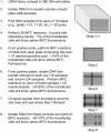
Flow Chart of cDNA Library Screening for Interacting Proteins. The steps in the screening process are presented, starting with DNA isolation from mixed colonies of plates to identification of individual cDNA clones encoding interacting proteins. For each step, a plasmid expressing an mRFP or mCherry reporter protein is cotransfected with the bait and cDNA library plasmids to evaluate transformation frequency (percentage of total cells that fluoresce red). Black boxes encircling plates, rows, columns, or individual colonies indicate which cDNA library plasmids are evaluated for each step of the process. The entire screening process takes 4 to 8 weeks.

References

    1. Anand A., Krichevsky A., Schornack S., Lahaye T., Tzfira T., Tang Y., Citovsky V., Mysore K.S. (2007). Arabidopsis VIRE2 INTERACTING PROTEIN2 is required for Agrobacterium T-DNA integration in plants. Plant Cell 19: 1695–1708 - PMC - PubMed
    1. Bakó L., Umeda M., Tiburcio A.F., Schell J., Koncz C. (2003). The VirD2 pilot protein of Agrobacterium-transferred DNA interacts with the TATA box-binding protein and a nuclear protein kinase in plants. Proc. Natl. Acad. Sci. USA 100: 10108–10113 - PMC - PubMed
    1. Ballas N., Citovsky V. (1997). Nuclear localization signal binding protein from Arabidopsis mediates nuclear import of Agrobacterium VirD2 protein. Proc. Natl. Acad. Sci. USA 94: 10723–10728 - PMC - PubMed
    1. Bartel P.L., Fields S. (1997). The Yeast Two-Hybrid System. (New York: Oxford University Press: ).
    1. Bhat R.A., Lahaye T., Panstruga R. (2006). The visible touch: in planta visualization of protein-protein interactions by fluorophore-based methods. Plant Methods 2: 12. - PMC - PubMed

Publication types

MeSH terms