. 2012 May 23;32(21):7301-10.
doi: 10.1523/JNEUROSCI.0185-12.2012.

In vivo visualization of delta opioid receptors upon physiological activation uncovers a distinct internalization profile

Affiliations

In vivo visualization of delta opioid receptors upon physiological activation uncovers a distinct internalization profile

Lauren Faget et al. J Neurosci. .

Abstract

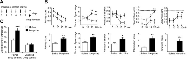
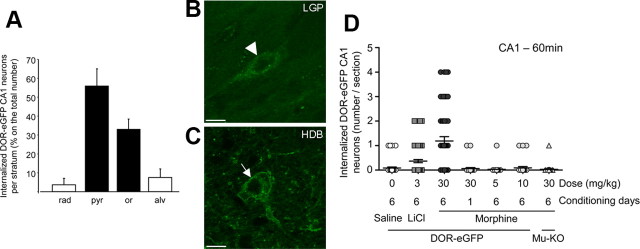
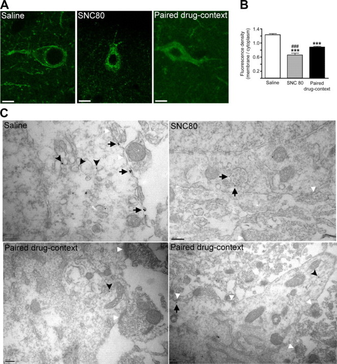
G-protein-coupled receptors (GPCRs) mediate numerous physiological functions and represent prime therapeutic targets. Receptor trafficking upon agonist stimulation is critical for GPCR function, but examining this process in vivo remains a true challenge. Using knock-in mice expressing functional fluorescent delta opioid receptors under the control of the endogenous promoter, we visualized in vivo internalization of this native GPCR upon physiological stimulation. We developed a paradigm in which animals were made dependent on morphine in a drug-paired context. When re-exposed to this context in a drug-free state, mice showed context-dependent withdrawal signs and activation of the hippocampus. Receptor internalization was transiently detected in a subset of CA1 neurons, uncovering regionally restricted opioid peptide release. Importantly, a pool of surface receptors always remained, which contrasts with the in vivo profile previously established for exogenous drug-induced internalization. Therefore, a distinct response is observed at the receptor level upon a physiological or pharmacological stimulation. Altogether, direct in vivo GPCR visualization enables mapping receptor stimulation promoted by a behavioral challenge and represents a powerful approach to study endogenous GPCR physiology.


Figures

Figure 1.
Figure 1.
Re-exposure to drug-paired context induces withdrawal signs. A, Drug-context pairing paradigm. DOR-eGFP mice received daily morphine (30 mg/kg, s.c.) or saline (NaCl 0.9%, s.c.) injections for 6 d, and were confined in a specific context for 60 min following injection. On the test day, mice received a saline injection and were re-exposed to the drug-associated context. Behavior was scored in 5 min bin periods for 20 min. B, Scores of the most significant behavioral signs on test day. Left, Activity (horizontal activity). Middle, Signs of discomfort (grooming, head shakes and wet dog shakes).Right, Vegetative signs (piloerection and chewing). Total scores (bottom) were compared using a Student's t test and time courses (top) were compared using a two-way ANOVA of repeated measures and a Newman–Keuls post hoc analysis. n = 12. C, The global behavioral score was significantly higher in morphine-treated mice submitted to the paired protocol (n = 12) compared with saline-treated mice (n = 12). No difference was observed between morphine-treated (n = 13) and saline-treated (n = 10) mice submitted to an unpaired protocol. Two-way ANOVA interaction of conditioning protocol (paired, unpaired) and drug treatment (morphine, saline): F(1,42) = 10.12; p = 0.0027. *p < 0.05; **p < 0.01; ***p < 0.001 morphine versus saline.
Figure 2.
Figure 2.
Context-induced withdrawal requires mu opioid receptor and compares to low-dose naloxone-precipitated withdrawal. A, Withdrawal global score in DOR-eGFP, wild-type, and MOR-KO mice re-exposed to the drug-paired context. Each global score was compared with the basal global score of their saline counterpart (100%). There is a general effect of the morphine treatment (F(3,37) = 10,879; p < 0.0001) but no difference is observed between morphine (n = 8) and saline (n = 6) mice deficient for mu opioid receptor submitted to the paired protocol (p = 0.5278). The global behavioral score is significantly higher in morphine-treated mice submitted to the paired protocol (n = 9) compared with saline-treated mice (n = 8) in wild-type animals (WT) (p = 0.0042) and in DOR-eGFP animals (n = 12) (p < 0.001). There is no difference between global scores of withdrawal from WT and DOR-eGFP mice (p = 0.2749). **p < 0.01; ***p < 0.001; one-way ANOVA, comparison with saline global score. B, Comparison between naloxone-precipitated and context-induced withdrawal after chronic morphine treatment. Pharmacological withdrawal precipitated by 0.3, 0.5, and 1 mg/kg naloxone induced significantly higher global behavioral scores in chronic morphine-treated mice (6 d, 30 mg/kg, s.c.) (n = 8) compared with control animals (n = 8) (effect of the treatment F(7,57) = 29.57; p < 0.001 and Tukey's post hoc analysis: p < 0.001). Mice chronically administered with morphine in the context (drug-context association protocol) also showed a higher global behavioral score than saline-injected animals (p = 0.018). This score was within the same range as scores observed for 0.3 mg/kg naloxone-precipitated withdrawal (dashed line) (p = 0.849). *p < 0.05; ***p < 0.001; one-way ANOVA, comparison with saline-treated mice 0-0 (pharmacological withdrawal); ###p < 0.001; one-way ANOVA, comparison with morphine-treated mice 30-0 (drug-context association protocol).
Figure 3.
Figure 3.
Re-exposure to drug-paired context induces c-fos expression in the hippocampus. A, Brightfield photomicrographs of hippocampal CA1 c-fos labeling in different paradigms. Top, Paired conditioning (saline and morphine). Bottom, Unpaired conditioning (saline and morphine). Scale bar, 250 μm. B, Number of c-fos-expressing cells per millimeter squared in the three main areas of the hippocampus in morphine- and saline-treated mice in the paired or unpaired protocols. Three-way ANOVA analysis revealed an interaction between region (DG, CA3, CA1), conditioning protocol (paired, unpaired), and drug treatment (morphine, saline): F(2,310) = 7.749; p < 0.001. Chronic morphine treatment (30 mg/kg) significantly increased the number of c-fos-positive cells in CA3 and CA1 regions re-exposed to the drug-paired context compared with saline-treated mice. No difference was observed between morphine- and saline-treated mice in the unpaired drug-context protocol. (saline n = 4; morphine n = 6). C, Chronic morphine treatment (30 mg/kg) also significantly increased the number of c-fos-positive cells in the BLA in animals re-exposed to the drug-paired context compared with saline-treated mice. D, Chronic morphine treatment (30 mg/kg) did not affect the number of c-fos-positive cells in the thalamic PVT used as a negative control region. **p < 0.01; ***p < 0.001 morphine versus saline; #p < 0.05; ###p < 0.001 paired morphine versus unpaired morphine.
Figure 4.
Figure 4.
DOR-eGFP-expressing neurons in the CA1 are GABAergic neurons mainly coexpressing parvalbumin. A, DOR-eGFP-expressing neurons of the CA1 are GABAergic neurons. GABAergic neurons are detected with an anti-GAD65/67 antibody and visualized using Alexa Fluor 594-conjugated secondary antibodies. DOR-eGFP green fluorescence is visible at cell surface (arrowhead). B, DOR-eGFP-expressing neurons of the stratum oriens and stratum pyramidale are mostly parvalbumin-positive GABAergic interneurons (Alexa Fluor 594 fluorescence). Rad, stratum radiatum; pyr, stratum pyramidale; or, stratum oriens; alv, stratum oriens alveus. Confocal fluorescence photomicrographs in 20-μm-thick brain sections. Scale bars: A, 10 μm; B, 50 μm.
Figure 5.
Figure 5.
DOR-eGFP internalization is mainly detectable in the dorsal CA1 area of the hippocampus upon re-exposure to the drug-paired context. Time course of DOR-eGFP internalization in the dorsal CA1 (A), CA3 (B), and DG (C) of the hippocampus on the test day after chronic morphine or saline treatment. The maximal number of DOR-eGFP-internalized neurons per section was observed 60 min after re-exposure to the drug-paired context in morphine-treated mice (10–12 sections/animal; n = 4). D, DOR-eGFP internalization in morphine-treated mice was higher in the CA1 compared with the CA3 and DG areas after re-exposure for 60 min to the drug-paired context (10–12 sections/animal; n = 4) Dark circles, morphine-treated mice; open circles, saline-treated mice.
Figure 6.
Figure 6.
DOR-eGFP internalization in the CA1 parvalbumin-positive GABAergic neurons is specific to re-exposure to the drug-paired context. A, There are 88.9 ± 3.8% of the internalized DOR-eGFP neurons located in the stratum pyramidale (pyr) and stratum oriens (or) (dark bars; 10–12 sections/animal; n = 4). Rad, stratum radiatum; pyr, stratum pyramidale; or, stratum oriens; alv, stratum oriens alveus. B, C, Representative images from confocal fluorescence micrographs of DOR-eGFP-expressing neurons in control regions. B, DOR-eGFP was internalized in almost all neurons of the LGP and in all of the tested conditions including morphine, saline, and paired and unpaired groups (arrowhead). C, In the HDB, DOR-eGFP did not internalize and fluorescence remained at the cell surface (arrow). Scale bar, 10 μm. D, DOR-eGFP internalization in the dorsal CA1 after re-exposure to the context (60 min) was higher in morphine-treated mice submitted to the drug-paired context compared with all other control conditions (10–12 sections/animal; n = 4). Control groups included saline-conditioned (open circles) and LiCl-conditioned animals (gray squares), and one day-conditioning or lower morphine doses (gray circles) as well as mu-opioid receptor knock-out mice (Mu-KO) (gray triangles).
Figure 7.
Figure 7.
DOR-eGFP is phosphorylated upon re-exposure to the drug-paired context. Representative confocal micrographs showing DOR-eGFP internalization (green signal) and delta opioid receptor phosphorylation (red signal) as revealed with an anti-phospho-delta Serine363 antibody detected with an Alexa Fluor 594-conjugated secondary antibody. Nuclei were stained with DAPI (blue). Context-exposed morphine-treated mice (60 min) resulted in colocalized intracellular DOR-eGFP and phospho-delta 363S antibody staining (white arrows). A, low magnification; B, high magnification. Scale bar, 10 μm.
Figure 8.
Figure 8.
DOR-eGFP internalization differs from the classical drug-induced internalization pattern. A, Representative confocal fluorescence micrographs of DOR-eGFP-expressing neurons in 20-μm-thick brain sections. In saline control mice (saline), DOR-eGFP fluorescence remained located at the membrane (left). In mice treated with the delta opioid agonist SNC80 (SNC80; 10 mg/kg, s.c., 30 min), DOR-eGFP was entirely located in intracellular vesicles forming the classically described endosomal punctuate pattern (middle). In morphine-treated mice re-exposed to the context (paired drug-context, 60 min), DOR-eGFP fluorescence increased inside the cell and appeared as an almost homogenous diffuse staining. Some fluorescence remained present at the plasma membrane (right). Scale bar, 10 μm. B, Quantification of DOR-eGFP internalization expressed as a ratio of membrane-associated versus cytoplasmic fluorescence densities (n = 11). ***p < 0.001 compared with saline; ###p < 0.001 SNC80-treated versus paired drug-context. C, Correlative light-electron microscopy. Images are representative micrographs showing eGFP immunolabeling in DOR-eGFP-expressing neurons. In saline-treated animals (saline), DOR-eGFP was located at the cell surface (black arrows) and in the Golgi apparatus (black arrowheads). In SNC80-treated animals (SNC80; 10 mg/kg s.c., 60 min) DOR-eGFP was present on multivesicular bodies (black arrows). Sixty minutes after re-exposure to the context previously associated with morphine (paired drug-context), DOR-eGFP was identified on intracellular vesicles close to synapses in both cell bodies and dendrites of neurons (black arrowheads) and was also present at the cell surface (black arrow). Scale bars, 200 nm, except bottom right panel, 500 nm. White arrowheads: synaptic boutons.
Figure 9.
Figure 9.
Enkephalin release is responsible for DOR-eGFP internalization. The enkephalinase inhibitor RB101 (60 mg/kg, i.p.) was administered immediately before re-exposure to the drug-paired context on the test day. A, In saline-treated mice, RB101 did not modify DOR-eGFP cell membrane distribution. B, In morphine-treated mice receiving saline solution, DOR-eGFP intracellular signal intensified as previously observed (Fig. 8). C, D, In morphine-treated mice that received RB101, DOR-eGFP fluorescence was still present at the membrane, but was also detected in intracellular vesicles similar to those observed after SNC80 treatment (arrows). Representative DOR-eGFP-expressing neurons in the CA1 stratum pyramidale (C) and stratum oriens (D) are shown. Nuclei were stained with DAPI (blue). Scale bar, 10 μm.

Similar articles

Cited by

References

    1. Berrendero F, Castañé A, Ledent C, Parmentier M, Maldonado R, Valverde O. Increase of morphine withdrawal in mice lacking A2a receptors and no changes in CB1/A2a double knockout mice. Eur J Neurosci. 2003;17:315–324. - PubMed
    1. Ciccocioppo R, Martin-Fardon R, Weiss F. Effect of selective blockade of mu(1) or delta opioid receptors on reinstatement of alcohol-seeking behavior by drug-associated stimuli in rats. Neuropsychopharmacology. 2002;27:391–399. - PubMed
    1. David V, Matifas A, Gavello-Baudy S, Decorte L, Kieffer BL, Cazala P. Brain regional Fos expression elicited by the activation of mu- but not delta-opioid receptors of the ventral tegmental area: evidence for an implication of the ventral thalamus in opiate reward. Neuropsychopharmacology. 2008;33:1746–1759. - PubMed
    1. Decossas M, Bloch B, Bernard V. Trafficking of the muscarinic m2 autoreceptor in cholinergic basalocortical neurons in vivo: differential regulation of plasma membrane receptor availability and intraneuronal localization in acetylcholinesterase-deficient and -inhibited mice. J Comp Neurol. 2003;462:302–314. - PubMed
    1. Enquist J, Kim J, Bartlett S, Ferwerda M, Whistler JL. A novel knock-in mouse reveals mechanistically distinct forms of morphine tolerance. J Pharmacol Exp Ther. 2011;338:633–640. - PMC - PubMed

Publication types

LinkOut - more resources