Review
. 2012 Jun;263(3):633-43.
doi: 10.1148/radiol.12102394.

Molecular body imaging: MR imaging, CT, and US. part I. principles

Affiliations
Review

Molecular body imaging: MR imaging, CT, and US. part I. principles

Moritz F Kircher et al. Radiology. 2012 Jun.

Abstract

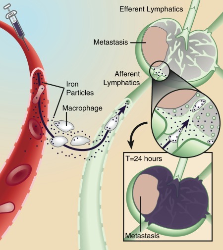
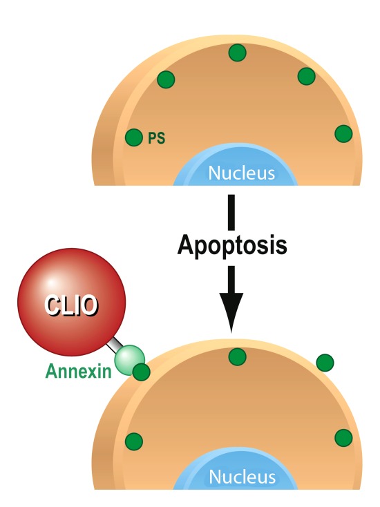
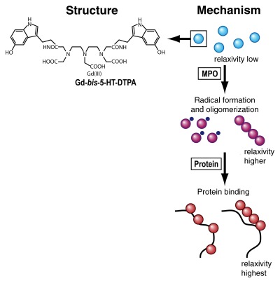
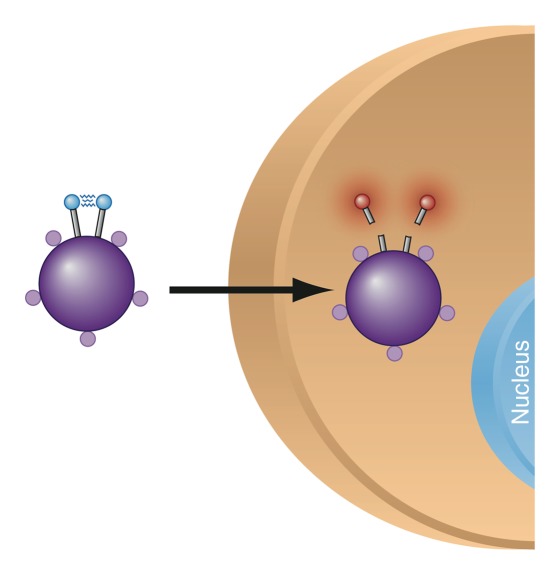
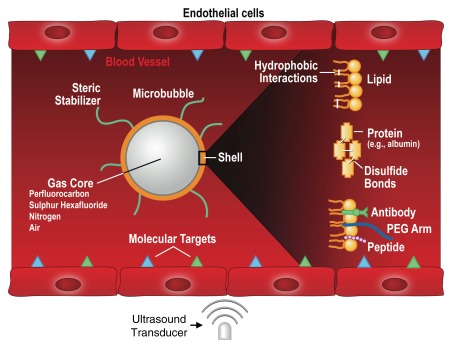
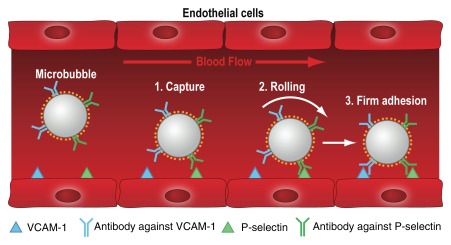
Molecular imaging, generally defined as noninvasive imaging of cellular and subcellular events, has gained tremendous depth and breadth as a research and clinical discipline in recent years. The coalescence of major advances in engineering, molecular biology, chemistry, immunology, and genetics has fueled multi- and interdisciplinary innovations with the goal of driving clinical noninvasive imaging strategies that will ultimately allow disease identification, risk stratification, and monitoring of therapy effects with unparalleled sensitivity and specificity. Techniques that allow imaging of molecular and cellular events facilitate and go hand in hand with the development of molecular therapies, offering promise for successfully combining imaging with therapy. While traditionally nuclear medicine imaging techniques, in particular positron emission tomography (PET), PET combined with computed tomography (CT), and single photon emission computed tomography, have been the molecular imaging methods most familiar to clinicians, great advances have recently been made in developing imaging techniques that utilize magnetic resonance (MR), optical, CT, and ultrasonographic (US) imaging. In the first part of this review series, we present an overview of the principles of MR imaging-, CT-, and US-based molecular imaging strategies.


Figures

Figure 1:
Figure 1:
Mechanism of action for lymphotropic superparamagnetic nanoparticles, one of the first clinically used cellular MR contrast agents. Systemically injected particles gain access to the interstitium and are drained through lymphatic vessels. In a normal lymph node, iron oxide nanoparticles are taken up by phagocytic cells, which cause the lymph node to become dark on T2-weighted images due to susceptibility artifacts from iron. If a lymph node is partially or fully replaced by metastatic cells, fewer nanoparticles are retained in the lymph node, which therefore remains bright on T2-weighted images. (Adapted and reprinted, with permission, from reference 36)
Figure 2:
Figure 2:
Mechanism of action for targeted MR molecular contrast agent for apoptosis imaging. As one of the early changes that occur when cells undergo apoptosis, phosphatidylserines (PS, green circles) become externalized on the cell membrane. Annexin V, conjugated to an iron oxide nanoparticle (CLIO), recognizes and binds to the externalized phosphatidylserines, leading to accumulation of the contrast agent; there is consecutive detectable signal intensity change on MR images.
Figure 3:
Figure 3:
Chemical structure of activatable MR molecular probe (left) and mechanism of action (right). The probe, gadolinium-5-hydroxytryptamide–diethylene triamine pentaacetic acid (Gd-bis-5-HT-DTPA) has a relatively low relaxivity (low visibility on MR images) in its native state (blue spheres). When the probe reaches an environment rich in myeloperoxidase (MPO), an enzyme secreted by white blood cells in inflammation, its monomers undergo rapid condensation into paramagnetic oligomers (purple spheres), leading to an increase in atomic relaxivity. This change in relaxivity is due to modulation of the rotational correlation time τr. Likewise, the relaxivity and therefore the MR signal increase even further when the probe binds to proteins (red spheres). (Adapted and reprinted, with permission, from reference .)
Figure 4a:
Figure 4a:
(a) Mechanism of action for dual-modality, activatable, magneto-optical molecular imaging MR contrast agent, which consists of a core of a dextran-coated iron oxide nanoparticle, onto which fluorochrome A (small purple spheres) is conjugated. Also attached to its surface are peptides carrying a second fluorochrome B (blue spheres) that can be cleaved by a specific enzyme. When the contrast agent enters an environment where the specific enzyme is present (eg, cathepsin B, which is overexpressed in breast cancer), the fluorochrome-carrying peptides are cleaved and thus fluorochrome B detaches. The increased distance of the fluorochromes causes dequenching, resulting in increased fluorescence signal. The intensity of fluorochrome A remains the same. (b–d) Illustration of fluorescence ratio method. Fluorescence is normally depth dependent and difficult to quantify. However, if two fluorochromes are used, where one is nonactivatable and serves as an internal standard, correction for depth is possible. While the signal from both fluorochromes used here (Cy5.5 and Cy7) decreases with increasing depth, the ratio of the two signal intensities remains the same (c, d). (Images bd reprinted, with permission, from reference .)
Figure 4b:
Figure 4b:
(a) Mechanism of action for dual-modality, activatable, magneto-optical molecular imaging MR contrast agent, which consists of a core of a dextran-coated iron oxide nanoparticle, onto which fluorochrome A (small purple spheres) is conjugated. Also attached to its surface are peptides carrying a second fluorochrome B (blue spheres) that can be cleaved by a specific enzyme. When the contrast agent enters an environment where the specific enzyme is present (eg, cathepsin B, which is overexpressed in breast cancer), the fluorochrome-carrying peptides are cleaved and thus fluorochrome B detaches. The increased distance of the fluorochromes causes dequenching, resulting in increased fluorescence signal. The intensity of fluorochrome A remains the same. (b–d) Illustration of fluorescence ratio method. Fluorescence is normally depth dependent and difficult to quantify. However, if two fluorochromes are used, where one is nonactivatable and serves as an internal standard, correction for depth is possible. While the signal from both fluorochromes used here (Cy5.5 and Cy7) decreases with increasing depth, the ratio of the two signal intensities remains the same (c, d). (Images bd reprinted, with permission, from reference .)
Figure 4c:
Figure 4c:
(a) Mechanism of action for dual-modality, activatable, magneto-optical molecular imaging MR contrast agent, which consists of a core of a dextran-coated iron oxide nanoparticle, onto which fluorochrome A (small purple spheres) is conjugated. Also attached to its surface are peptides carrying a second fluorochrome B (blue spheres) that can be cleaved by a specific enzyme. When the contrast agent enters an environment where the specific enzyme is present (eg, cathepsin B, which is overexpressed in breast cancer), the fluorochrome-carrying peptides are cleaved and thus fluorochrome B detaches. The increased distance of the fluorochromes causes dequenching, resulting in increased fluorescence signal. The intensity of fluorochrome A remains the same. (b–d) Illustration of fluorescence ratio method. Fluorescence is normally depth dependent and difficult to quantify. However, if two fluorochromes are used, where one is nonactivatable and serves as an internal standard, correction for depth is possible. While the signal from both fluorochromes used here (Cy5.5 and Cy7) decreases with increasing depth, the ratio of the two signal intensities remains the same (c, d). (Images bd reprinted, with permission, from reference .)
Figure 4d:
Figure 4d:
(a) Mechanism of action for dual-modality, activatable, magneto-optical molecular imaging MR contrast agent, which consists of a core of a dextran-coated iron oxide nanoparticle, onto which fluorochrome A (small purple spheres) is conjugated. Also attached to its surface are peptides carrying a second fluorochrome B (blue spheres) that can be cleaved by a specific enzyme. When the contrast agent enters an environment where the specific enzyme is present (eg, cathepsin B, which is overexpressed in breast cancer), the fluorochrome-carrying peptides are cleaved and thus fluorochrome B detaches. The increased distance of the fluorochromes causes dequenching, resulting in increased fluorescence signal. The intensity of fluorochrome A remains the same. (b–d) Illustration of fluorescence ratio method. Fluorescence is normally depth dependent and difficult to quantify. However, if two fluorochromes are used, where one is nonactivatable and serves as an internal standard, correction for depth is possible. While the signal from both fluorochromes used here (Cy5.5 and Cy7) decreases with increasing depth, the ratio of the two signal intensities remains the same (c, d). (Images bd reprinted, with permission, from reference .)
Figure 5a:
Figure 5a:
Mechanism of action for MR hyperpolarization. The fundamental principle of MR imaging is based on the interaction of atomic nuclei with an external magnetic field. (a) Nuclei can orient themselves in two possible directions: parallel (“spin up,” α) or antiparallel (“spin-down,” β) to the external field (B0). If the two populations are equal, their magnetic moments cancel, resulting in no MR signal. In thermal equilibrium, there are a very small number of unequally oriented spins (∼ 1 in 105). Only this small number of spins can contribute to the MR signal, resulting in a low sensitivity. (b) The idea of hyperpolarization is to create an artificial nonequilibrium of spins. This means that the number of unequally oriented spins will be increased by a factor of up to 100 000, which therefore results in a much higher MR signal.
Figure 5b:
Figure 5b:
Mechanism of action for MR hyperpolarization. The fundamental principle of MR imaging is based on the interaction of atomic nuclei with an external magnetic field. (a) Nuclei can orient themselves in two possible directions: parallel (“spin up,” α) or antiparallel (“spin-down,” β) to the external field (B0). If the two populations are equal, their magnetic moments cancel, resulting in no MR signal. In thermal equilibrium, there are a very small number of unequally oriented spins (∼ 1 in 105). Only this small number of spins can contribute to the MR signal, resulting in a low sensitivity. (b) The idea of hyperpolarization is to create an artificial nonequilibrium of spins. This means that the number of unequally oriented spins will be increased by a factor of up to 100 000, which therefore results in a much higher MR signal.
Figure 6a:
Figure 6a:
Principle of a nanoparticulate CT contrast agent. (a) Schematic representation of the contrast-generating iodinated compound of the nanoparticle CT contrast agent N1177 containing three iodine (I) atoms (red). (b) Electron microscopy image of N1177. The nanoparticles are suspensions of crystalline iodinated compound, combined with biocompatible surfactants to prevent aggregation and stabilize nanoparticle size. Shown are electron-dense iodinated granules coated by polymers appearing as negative prints after staining with a solution of uranyl acetate. Nanoparticles have various sizes and shapes. Scale bar = 100 nm. (Adapted and reprinted, with permission, from reference .)
Figure 6b:
Figure 6b:
Principle of a nanoparticulate CT contrast agent. (a) Schematic representation of the contrast-generating iodinated compound of the nanoparticle CT contrast agent N1177 containing three iodine (I) atoms (red). (b) Electron microscopy image of N1177. The nanoparticles are suspensions of crystalline iodinated compound, combined with biocompatible surfactants to prevent aggregation and stabilize nanoparticle size. Shown are electron-dense iodinated granules coated by polymers appearing as negative prints after staining with a solution of uranyl acetate. Nanoparticles have various sizes and shapes. Scale bar = 100 nm. (Adapted and reprinted, with permission, from reference .)
Figure 7:
Figure 7:
Overview of different types of US contrast agents. Microbubbles are gas-liquid emulsions with a polyethylene glycol (PEG) polymer on their surface to prevent aggregation. Perfluorocarbon emulsion nanodroplets are liquid-liquid emulsions that can be vaporized into echogenic gas bubbles after administration of acoustic energy. Liposomes are phospholipid bilayers that can enclose air pockets for US imaging. Nanobubbles are gas-liquid emulsions that can fuse into echogenic microbubbles at the target site. Solid nanoparticles are solid amorphous substances with gas entrapped in their pores or fissures, which increases their echogenicity. (Adapted and reprinted, with permission, from reference .)
Figure 8:
Figure 8:
Mechanism of action for targeted US microbubbles. Targeted microbubbles consist of a gas core that can contain different types of gas (eg, perfluorocarbons or nitrogen) surrounded by a shell of, for example, phospholipids or proteins. The microbubble shell can be functionalized by attaching binding ligands such as antibodies or small peptides, which allows the microbubbles to accumulate at sites that overexpress molecular targets (blue and green triangles). Because the size of microbubbles is of several micrometers, they stay exclusively within the vascular compartment, which makes them most useful for imaging and quantification of molecular targets that are luminally overexpressed on vascular endothelial cells, such as markers of angiogenesis or inflammation. When exposed to an ultrasound field, the microbubbles oscillate and send out nonlinear acoustic signals that can be detected with currently clinically used US systems. Since the imaging signal from microbubbles is substantially higher than the signal from surrounding tissue, microbubble accumulation can be imaged with high signal-to-background ratio. PEG = polyethylene glycol. (Adapted and reprinted, with permission, from reference .)
Figure 9:
Figure 9:
Mechanism of action for dual-targeted microbubbles. Dual-targeted microbubbles can be designed to mimic the behavior of leukocytes in vivo. The diagram shows a dual-targeted microbubble targeted against both P-selectin and vascular cell adhesion molecule-1 (VCAM-1), simulating vascular attachment of a leukocyte at sites of inflammation by first interacting with P-selectin and then firmly attaching via VCAM1. (Adapted and reprinted, with permission, from reference .)

References

    1. Weissleder R, Pittet MJ. Imaging in the era of molecular oncology. Nature 2008;452(7187):580–589 - PMC - PubMed
    1. Mazaheri Y, Shukla-Dave A, Muellner A, Hricak H. MRI of the prostate: clinical relevance and emerging applications. J Magn Reson Imaging 2011;33(2):258–274 - PubMed
    1. Vargas HA, Akin O, Franiel T, et al. Diffusion-weighted endorectal MR imaging at 3 T for prostate cancer: tumor detection and assessment of aggressiveness. Radiology 2011;259(3):775–784 - PMC - PubMed
    1. Biswal S, Resnick DL, Hoffman JM, Gambhir SS. Molecular imaging: integration of molecular imaging into the musculoskeletal imaging practice. Radiology 2007;244(3):651–671 - PubMed
    1. Pysz MA, Willmann JK. Targeted contrast-enhanced ultrasound: an emerging technology in abdominal and pelvic imaging. Gastroenterology 2011;140(3):785–790 - PMC - PubMed

Publication types

LinkOut - more resources