. 2012 May 23;4(135):135ra66.
doi: 10.1126/scitranslmed.3003759.

PTH/PTHrP and vitamin D control antimicrobial peptide expression and susceptibility to bacterial skin infection

Affiliations

PTH/PTHrP and vitamin D control antimicrobial peptide expression and susceptibility to bacterial skin infection

Beda Muehleisen et al. Sci Transl Med. .

Abstract

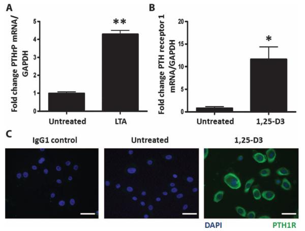
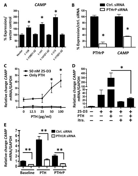
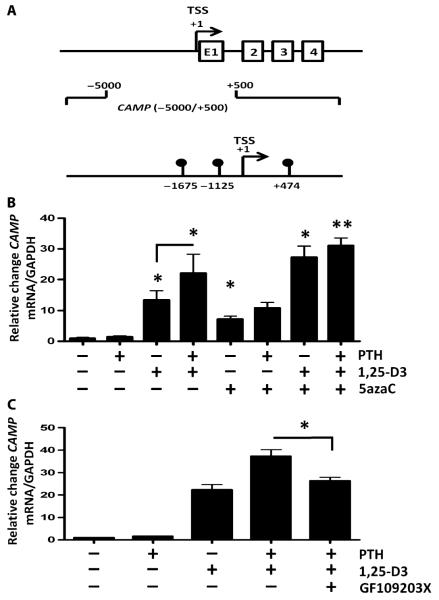
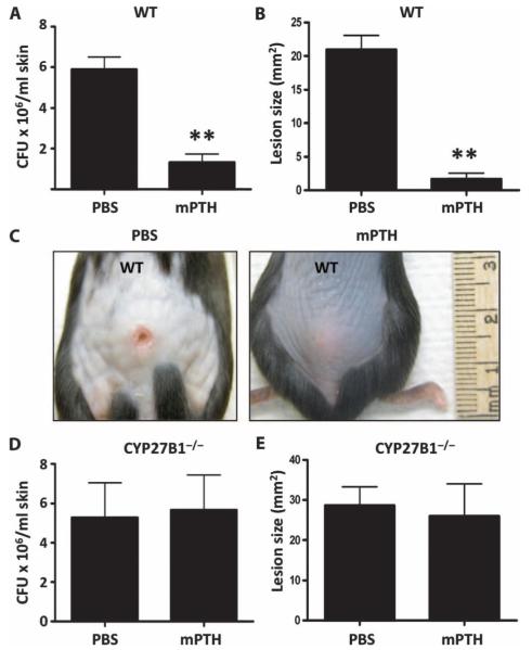
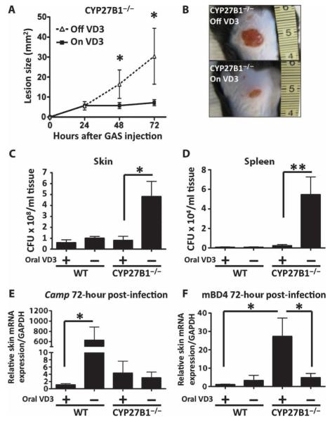
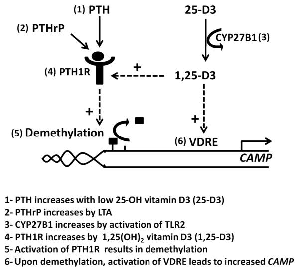
The production of antimicrobial peptides is essential for protection against a wide variety of microbial pathogens and plays an important role in the pathogenesis of several diseases. The mechanisms responsible for expression of antimicrobial peptides are incompletely understood, but a role for vitamin D as a transcriptional inducer of the antimicrobial peptide cathelicidin has been proposed. We show that 1,25-dihydroxyvitamin D(3) (1,25-D3) acts together with parathyroid hormone (PTH), or the shared amino-terminal domain of PTH-related peptide (PTHrP), to synergistically increase cathelicidin and immune defense. Administration of PTH to mouse skin decreased susceptibility to skin infection by group A Streptococcus. Mice on dietary vitamin D(3) restriction that responded with an elevation in PTH have an increased risk of infection if they lack 1,25-D3. These results identify PTH/PTHrP as a variable that serves to compensate for inadequate vitamin D during activation of antimicrobial peptide production.


Figures

Fig. 1
Fig. 1
LTA induces PTHrP and 1,25-D3 induces its receptor, PTH1R, in keratinocytes. (A) Expression of PTHrP mRNA in normal human epidermal keratinocytes 24 hours after treatment with the TLR2 agonist LTA (10 μg/ml). (B) Expression of PTH1R mRNA in normal human epidermal keratinocytes treated with 1,25-D3 (10−7 M) for 24 hours. (C) Immunofluorescence staining of keratinocytes using IgG1 control antibody (left panel) or anti-PTH1R antibody (green) on untreated (middle panel) or 1,25-D3–treated cells (right panel). Blue, staining of nuclei with 4′,6-diamidino-2-phenylindole (DAPI). Scale bars, 20 μm. Data are means ± SEM of triplicate independent cultures and are representative of two independent experiments. *P < 0.05, **P < 0.01 by unpaired t test. GAPDH, glyceraldehyde-3-phosphate dehydrogenase.
Fig. 2
Fig. 2
PTHrP and PTH cooperate with vitamin D3 to induce cathelicidin antimicrobial peptide expression. (A) Cathelicidin (CAMP) mRNA expression in HaCaT keratinocytes transfected for 48 hours with different PTHrP expression plasmids. N-terminal amino acids encoded by each construct are designated. Constructs labeled D1-32 lack amino acids 1 to 32 of PTHrP and were inactive. Data are means ± SEM of triplicate independent cultures and are representative of two independent experiments. *P < 0.05 by unpaired t test. (B) HaCaT keratinocytes transfected with control siRNA (black bars) or PTHrP siRNA duplexes (white bars) and grown for 48 hours. PTHrP was measured in conditioned media by immunoassay, and cathelicidin (CAMP) mRNA was measured in cell extracts by qPCR. Data are means ± SEM of triplicate independent cultures and are representative of two independent experiments. *P < 0.05 versus vector control (unpaired t test). (C) CAMP mRNA expression in normal human epidermal keratinocytes after treatment with PTH (0 to 100 pg/ml) alone or cotreated with 25-D3 (50 nM). *P < 0.05 by Kruskal-Wallis with Dunn’s post hoc nonparametric test. Data are means ± SEM of triplicate independent cultures and are representative of three independent experiments. (D) Normal human epidermal keratinocyte CAMP mRNA expression after pretreatment with the CYP27B1 inhibitor itraconazole (itra., 10−7 M) for 1 hour before pretreatment with 25-D3 (50 nM) for 24 hours and PTH (10−11 M) for another 24 hours. *P < 0.05 by unpaired t test. Data are means ± SEM of triplicate independent cultures and are representative of three independent experiments. (E) Normal human epidermal keratinocyte CAMP mRNA expression after knockdown using PTH1R siRNA (open bars) or control siRNA (Ctrl. siRNA, solid bars) at baseline (left) or after pretreatment for 24 hours with PTH (middle, 10−11 M) or PTHrP (right, 10−11 M). Data are means ± SEM of triplicate independent cultures and are representative of two independent experiments. **P < 0.01 by Mann-Whitney U test.
Fig. 3
Fig. 3
CAMP expression by PTH/PTHrP involves DNA methylation. (A) Whole-genome MeDIP of primary human keratinocytes revealed three methylated DNA sites within −5000/+500 bp of the CAMP gene TSS at −1675, −1125, and +474 bp. Shown are exons 1 to 4 (E1, 2, 3, 4) of the cathelicidin gene. (B) CAMP mRNA expression in normal human epidermal keratinocytes after treatment with PTH (10−11 M), 1,25-D3 (10−7 M), and the DNA methyltransferase inhibitor 5azaC (10−6 M). (C) CAMP mRNA expression after treatment with PTH (10−11 M), 1,25-D3 (10−7 M), and the PKC inhibitor GF109203X (5 μM) for 24 hours. Results are means ± SEM of triplicate independent cultures and are representative of three independent experiments. *P < 0.05, **P < 0.01 by Mann-Whitney U test.
Fig. 4
Fig. 4
PTH in the presence of vitamin D3 enhances protection against invasive bacterial skin infections in vivo. Wild-type (WT) mice and mice lacking the 1α-hydroxylase enzyme (CYP27B1−/−) were injected subcutaneously with mPTH(1–34) peptide (80 μg/kg) or PBS 24 hours before challenge with 107 CFU of group A Streptococcus NZ131. (A) Number of group A Streptococcus CFU retrieved from homogenized infected WT mouse skin after 72 hours. (B) Group A Streptococcus lesion size (mm2) in WT mice 72 hours after infection. **P < 0.01 by unpaired t test. (C) A representative photograph of group A Streptococcus lesion size in WT mice injected with mPTH or PBS at 72 hours. (D) Number of group A Streptococcus CFU retrieved from homogenized infected CYP27B1−/− mouse skin after 72 hours. (E) Group A Streptococcus lesion size (mm2) in CYP27B1−/− mice 72 hours after infection. All data are means ± SEM from six mice per group and are representative of three independent experiments.
Fig. 5
Fig. 5
Dietary vitamin D3 restriction in CYP27B1−/− mice increases susceptibility to group A Streptococcus infection. CYP27B1−/− mice were injected subcutaneously with 107 CFU of group A Streptococcus NZ131 after having been on a diet either containing vitamin D3 (2.2 IU/g) (“On VD3” or “+”) or not containing any vitamin D (“Off VD3” or “−”) for 4 weeks. (A) Lesion size of group A Streptococcus (GAS) skin infections (mm2) 24, 48, and 72 hours after injection. All data are means ± SEM from six mice per group and are representative of three independent experiments. *P < 0.05 by ANOVA with Bonferroni’s posttest. (B) A representative photograph of group A Streptococcus lesion size at 72 hours is shown. (C and D) Number of group A Streptococcus NZ131 CFU retrieved from homogenized skin (C) and spleen (D) 72 hours after infection. (E and F) Cathelicidin (Camp) (E) and mBD4 (F) mRNA expression in mouse skin 72 hours after group A Streptococcus injection. *P < 0.05, **P < 0.01 by Mann-Whitney U test.
Fig. 6
Fig. 6
Schematic showing how cathelicidin expression, PTH/PTHrP, and vitamin D may be connected. The human cathelicidin gene CAMP is induced by vitamin D, and its expression is further enhanced by the action of PTH/PTHrP. (1) PTH levels are elevated in response to low 25-D3 such as occurs with limited vitamin D intake. (2) PTHrP is increased locally in keratinocytes by the TLR2 agonist LTA. (3) The most active metabolite of vitamin D, 1,25-D3, is increased when TLR2 activation induces CYP27B1 to enhance conversion of 25-D3 to 1,25-D3. (4) 1,25-D3 enhances expression of PTH1R. (5) Activation of PTH1R results in demethylation of the CAMP promoter, thus enhancing expression of cathelicidin. (6) CAMP contains a functional vitamin D response element that induces its expression. These interactions define a potential mechanism to compensate for low serum 25-D3 by increasing the amount of PTH available for activation of CAMP, and also show how recognition of bacterial products can amplify cathelicidin expression by increasing both local 1,25-D3 and PTH1R.

Similar articles

Cited by

References

    1. Lai Y, Gallo RL. AMPed up immunity: How antimicrobial peptides have multiple roles in immune defense. Trends Immunol. 2009;30:131–141. - PMC - PubMed
    1. Braff MH, Bardan A, Nizet V, Gallo RL. Cutaneous defense mechanisms by antimicrobial peptides. J. Invest. Dermatol. 2005;125:9–13. - PubMed
    1. Lai Y, Cogen AL, Radek KA, Park HJ, Macleod DT, Leichtle A, Ryan AF, Di Nardo A, Gallo RL. Activation of TLR2 by a small molecule produced by Staphylococcus epidermidis increases antimicrobial defense against bacterial skin infections. J. Invest. Dermatol. 2010;130:2211–2221. - PMC - PubMed
    1. Ong PY, Ohtake T, Brandt C, Strickland I, Boguniewicz M, Ganz T, Gallo RL, Leung DY. Endogenous antimicrobial peptides and skin infections in atopic dermatitis. N. Engl. J. Med. 2002;347:1151–1160. - PubMed
    1. Schauber J, Dorschner RA, Coda AB, Büchau AS, Liu PT, Kiken D, Helfrich YR, Kang S, Elalieh HZ, Steinmeyer A, Zügel U, Bikle DD, Modlin RL, Gallo RL. Injury enhances TLR2 function and antimicrobial peptide expression through a vitamin D–dependent mechanism. J. Clin. Invest. 2007;117:803–811. - PMC - PubMed

Publication types

MeSH terms