. 2012 Aug;86(15):7976-87.
doi: 10.1128/JVI.00770-12. Epub 2012 May 23.

An Epstein-Barr Virus (EBV) mutant with enhanced BZLF1 expression causes lymphomas with abortive lytic EBV infection in a humanized mouse model

Affiliations

An Epstein-Barr Virus (EBV) mutant with enhanced BZLF1 expression causes lymphomas with abortive lytic EBV infection in a humanized mouse model

Shi-Dong Ma et al. J Virol. 2012 Aug.

Abstract

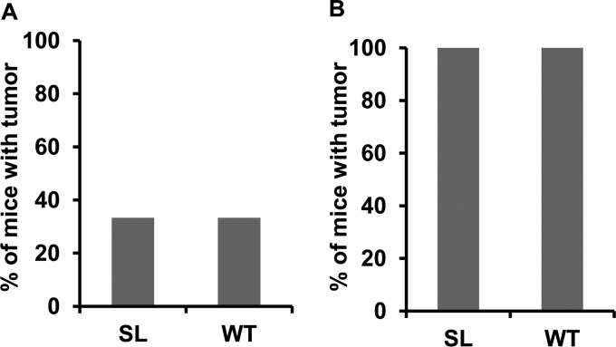
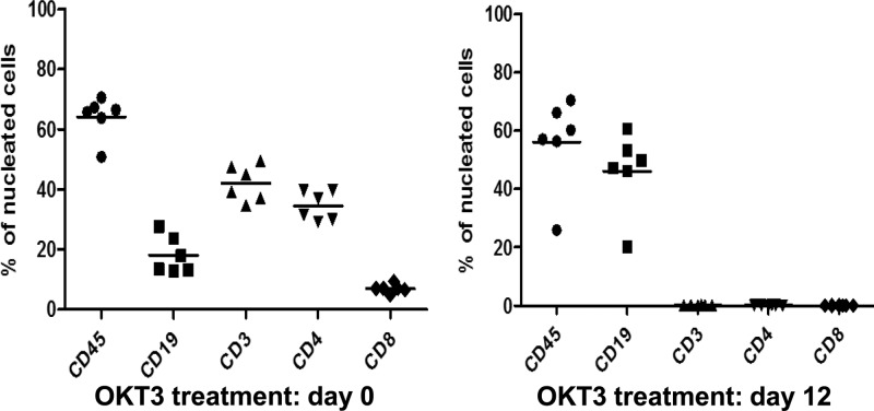
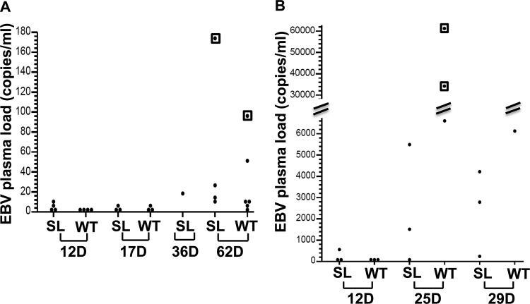
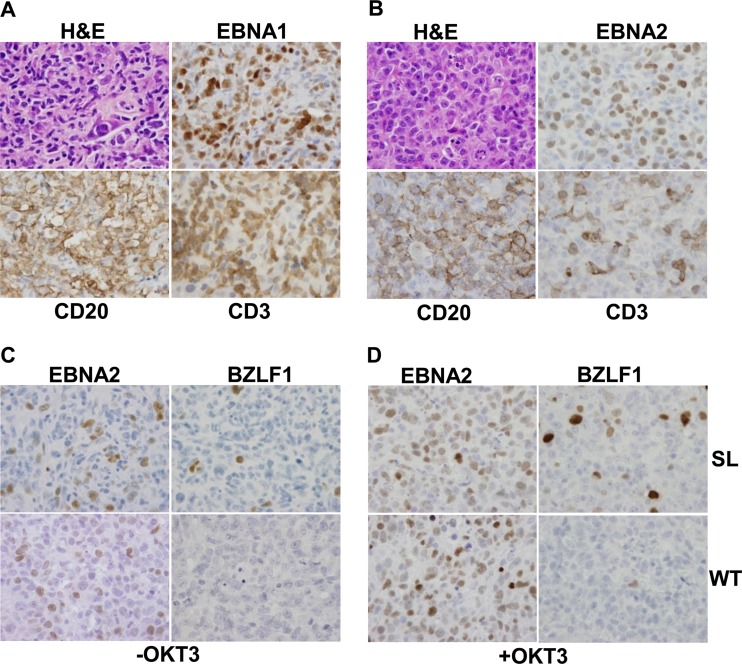
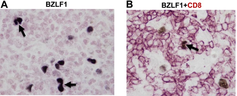
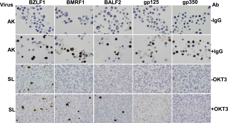
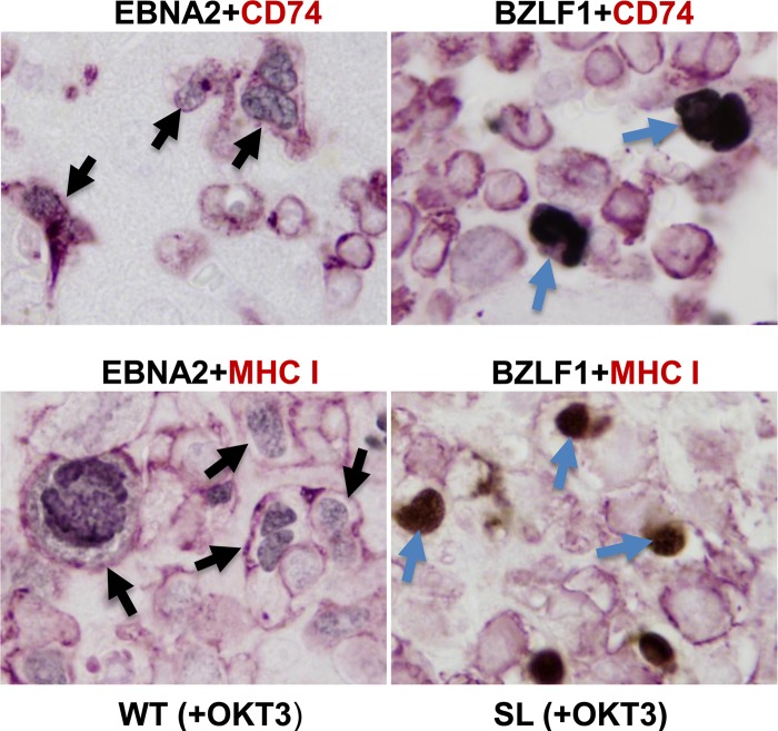
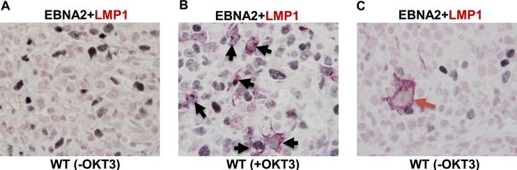
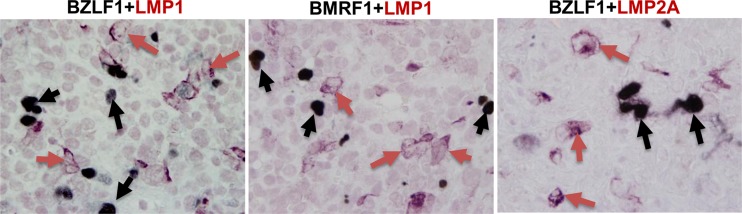
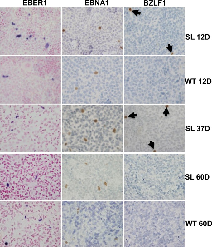
Immunosuppressed patients are at risk for developing Epstein-Barr Virus (EBV)-positive lymphomas that express the major EBV oncoprotein, LMP1. Although increasing evidence suggests that a small number of lytically infected cells may promote EBV-positive lymphomas, the impact of enhanced lytic gene expression on the ability of EBV to induce lymphomas is unclear. Here we have used immune-deficient mice, engrafted with human fetal hematopoietic stem cells and thymus and liver tissue, to compare lymphoma formation following infection with wild-type (WT) EBV versus infection with a "superlytic" (SL) mutant with enhanced BZLF1 (Z) expression. The same proportions (2/6) of the WT and SL virus-infected animals developed B-cell lymphomas by day 60 postinfection; the remainder of the animals had persistent tumor-free viral latency. In contrast, all WT and SL virus-infected animals treated with the OKT3 anti-CD3 antibody (which inhibits T-cell function) developed lymphomas by day 29. Lymphomas in OKT3-treated animals (in contrast to lymphomas in the untreated animals) contained many LMP1-expressing cells. The SL virus-infected lymphomas in both OKT3-treated and untreated animals contained many more Z-expressing cells (up to 30%) than the WT virus-infected lymphomas, but did not express late viral proteins and thus had an abortive lytic form of EBV infection. LMP1 and BMRF1 (an early lytic viral protein) were never coexpressed in the same cell, suggesting that LMP1 expression is incompatible with lytic viral reactivation. These results show that the SL mutant induces an "abortive" lytic infection in humanized mice that is compatible with continued cell growth and at least partially resistant to T-cell killing.


Figures

Fig 1
Fig 1
SL EBV mutant can induce lymphoma in hNSG(thy) mice. The percentage of mice developing EBV-positive tumors in SL versus WT virus-infected animals is shown. (A) Animals without OKT3 treatment; (B) animals treated with OKT3.
Fig 2
Fig 2
OKT3 treatment depleted peripheral blood human T cells in all treated mice. Peripheral blood was collected from hNSG(thy) mice at days 0 and 12 post-OKT3 treatment. Human CD45, CD19, CD3, CD4, and CD8 cell populations were detected by flow cytometry. Dead cells were excluded from the analysis. Results are presented as the percentage of positively staining cells with each antibody in comparison to the total leukocyte population.
Fig 3
Fig 3
The SL mutant and WT EBV-infected animals have similar viral loads in plasma, but OKT3 treatment increases viral load after EBV infection. qPCR was performed (using primers to detect the BamHIW repeat region of the EBV genome) on purified plasma DNA collected from SL mutant and WT EBV-infected animals at various days postinfection as indicated. (A) Viral load from animals without OKT3 treatment. The boxed dots indicate the two animals that developed tumors. (B) Viral load from animals treated with OKT3. The boxed dots indicate animals that were sacrificed at day 25 due to significant weight loss. All six animals had tumors.
Fig 4
Fig 4
SL virus induces DLBCLs with upregulated BZLF1 expression. (A) SL mutant EBV-induced DLBCL from a non-OKT3-treated animal was stained for H&E, EBV EBNA1 protein, CD20, and CD3 (100× magnification). Large atypical tumor cells showing CD20 membrane staining were infiltrated by numerous small CD3-staining T cells. (B) SL EBV-induced DLBCL from an OKT3-treated animal was stained for H&E, EBV EBNA1 protein, CD20, and CD3 (100× magnification). (C) EBNA2 and BZLF1 staining of DLBCLs in SL mutant EBV-infected (upper panel) and WT EBV-infected (lower panel) (100× magnification) animals not treated with OKT3. (D) EBNA2 and BZLF1 staining of DLBCLs in SL EBV-infected (upper panel) and WT EBV-infected (lower panel) (100× magnification) animals treated with OKT3.
Fig 5
Fig 5
BZLF1-expressing lymphoma cells can divide in hNSG(thy) mice. (A) BZLF1 staining of a DLBCL from an SL mutant EBV-infected animal (without OKT3 treatment). (B) Dual-color immunohistochemistry was performed using anti-BZLF1 (black) and anti-CD8 (pink) antibodies in an SL-EBV-infected animal (without OKT3 treatment). Dividing cells are indicated by black arrows (100× magnification).
Fig 6
Fig 6
The SL EBV mutant induces an abortive form of lytic EBV infection in lymphoma cells. Anti-BZLF1, anti-BMRF1, anti-BALF2, anti-gp125, and anti-350/220 staining was performed as indicated (100× magnification) to examine expression of immediate-early (BZLF1), early lytic (BMRF1 and BALF2), and late lytic (gp125 and gp350/220) viral proteins in SL EBV-infected animals. Formalin-fixed and paraffin-embedded Akata (AK) BL cells treated in vitro without or with anti-IgG pretreatment as indicated in the upper two panels served as the baseline and induced controls for lytic EBV protein expression, respectively. Abortive lytic infection is seen in animals with or without OKT3 treatment, as indicated in the lower two panels.
Fig 7
Fig 7
The EBV BMRF1 early lytic promoter is highly methylated in EBV-induced DLBCLs. DNA isolated from an SL mutant EBV-infected DLBCL without OKT3 treatment was bisulfite treated and sequenced. The black dots indicate the methylated CpGs, and white dots indicate the unmethylated CpGs in each sequenced clone. The numbers shown are relative to the BMRF1 transcript start site.
Fig 8
Fig 8
BZLF1-expressing cells have decreased expression of CD74 and MHC I. Dual-color immunohistochemistry was performed using anti-EBNA2 or anti-BZLF1 (black) and anti-CD74 or anti-MHC I (pink) antibodies on DLBCLs from OKT3-treated animals. EBNA2+ costaining was performed on a WT EBV-infected DLBCL, while BZLF1 costaining was performed on an SL mutant EBV-infected DLBCL. Examples of EBNA2 and CD74 or MHCI coexpression are indicated by black arrows; examples of BZLF1-positive cells lacking CD74 or MHCI are indicated by blue arrows.
Fig 9
Fig 9
OKT3 treatment greatly increases LMP1 and LMP2A expression in lymphoma cells. Dual-color immunohistochemistry was performed using anti-EBNA2 (black) and anti-LMP1 (pink) antibodies in DLBCLs from WT EBV-infected animals (A and C) and a DLBCL from a WT EBV-infected animal that had been treated with OKT3 antibody (B). Examples of EBNA2-positive cells costaining with LMP1 are indicated by black arrows in panel B. An example of a cell with type IIA latency (LMP1+ EBNA2) is indicated by a red arrow in panel C. All pictures are at 100× magnification.
Fig 10
Fig 10
BZLF1/BMRF1 and LMP1/LMP2A expression are mutually exclusive. Dual-color immunohistochemistry was performed using anti-BZLF1 or anti-BMRF1 (black) and anti-LMP1 or anti-LMP2A (pink) antibodies as indicated on SL EBV-infected DLBCLs from OKT3-treated animals. Cells expressing only BZLF1or BMRF1 are indicated by black arrows, while cells expressing only LMP1 or LMP2A are indicated by red arrows.
Fig 11
Fig 11
The SL mutant establishes tumor-free viral latency in some hNSG(thy) mice by day 60 postinfection. EBER1, EBNA1, and BZLF1 staining was performed on spleens of WT and SL mutant EBV-infected animals (not treated with OKT3) sacrificed at different days (D) postinfection as indicated (40× magnification). Examples of BZLF1-staining cells are indicated by arrows at the earlier time points, but at day 60, none of these tumor-free animals still had BZLF1-expressing cells.

Similar articles

Cited by

References

    1. Adamson AL, Kenney S. 2001. Epstein-Barr virus immediate-early protein BZLF1 is SUMO-1 modified and disrupts promyelocytic leukemia bodies. J. Virol. 75:2388–2399 - PMC - PubMed
    1. Adler B, et al. 2002. Control of Epstein-Barr virus reactivation by activated CD40 and viral latent membrane protein 1. Proc. Natl. Acad. Sci. U. S. A. 99:437–442 - PMC - PubMed
    1. Bailey SG, et al. 2009. Functional interaction between Epstein-Barr virus replication protein Zta and host DNA damage response protein 53BP1. J. Virol. 83:11116–11122 - PMC - PubMed
    1. Basha G, et al. 2012. A CD74-dependent MHC class I endolysosomal cross-presentation pathway. Nat. Immunol. 13:237–245 - PMC - PubMed
    1. Bhende PM, Dickerson SJ, Sun X, Feng WH, Kenney SC. 2007. X-box-binding protein 1 activates lytic Epstein-Barr virus gene expression in combination with protein kinase D. J. Virol. 81:7363–7370 - PMC - PubMed

Publication types

MeSH terms

Substances