. 2012 Aug;86(15):8185-97.
doi: 10.1128/JVI.07240-11. Epub 2012 May 23.

Arenavirus nucleoproteins prevent activation of nuclear factor kappa B

Affiliations

Arenavirus nucleoproteins prevent activation of nuclear factor kappa B

W W Shanaka I Rodrigo et al. J Virol. 2012 Aug.

Abstract

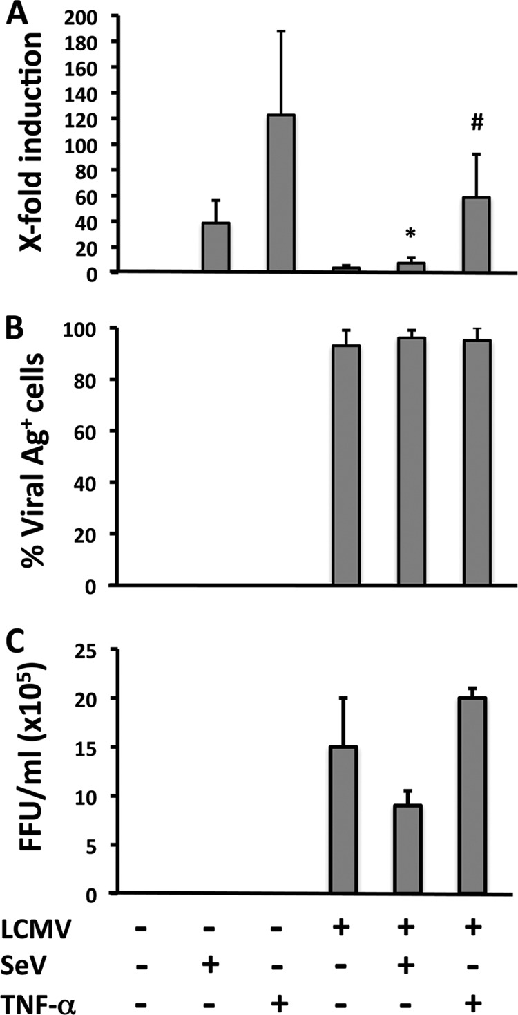
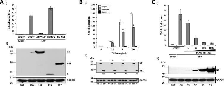
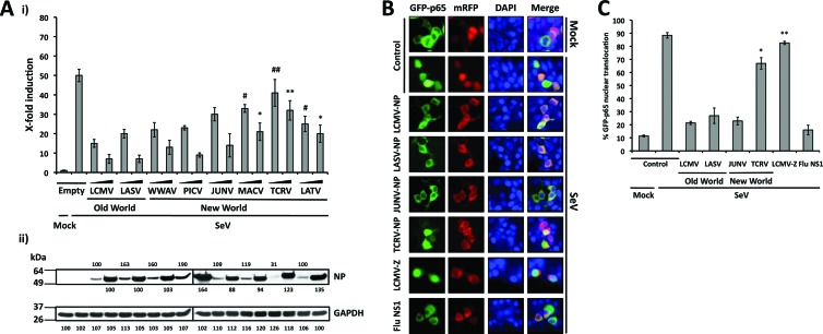
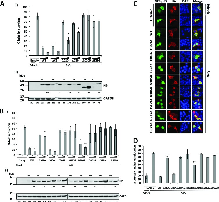
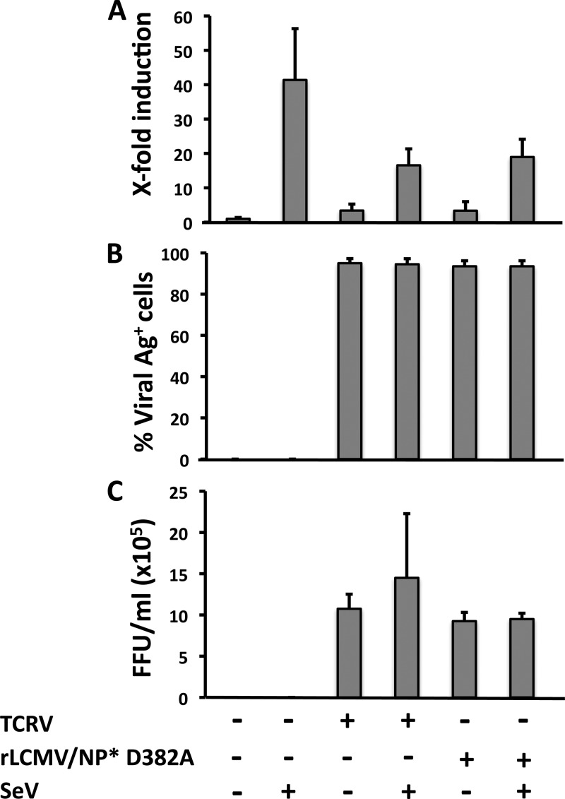
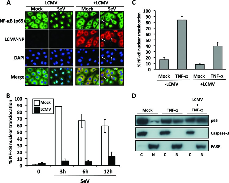
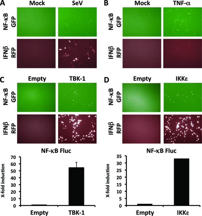
Arenaviruses include several causative agents of hemorrhagic fever (HF) disease in humans that are associated with high morbidity and significant mortality. Morbidity and lethality associated with HF arenaviruses are believed to involve the dysregulation of the host innate immune and inflammatory responses that leads to impaired development of protective and efficient immunity. The molecular mechanisms underlying this dysregulation are not completely understood, but it is suggested that viral infection leads to disruption of early host defenses and contributes to arenavirus pathogenesis in humans. We demonstrate in the accompanying paper that the prototype member in the family, lymphocytic choriomeningitis virus (LCMV), disables the host innate defense by interfering with type I interferon (IFN-I) production through inhibition of the interferon regulatory factor 3 (IRF3) activation pathway and that the viral nucleoprotein (NP) alone is responsible for this inhibitory effect (C. Pythoud, W. W. Rodrigo, G. Pasqual, S. Rothenberger, L. Martínez-Sobrido, J. C. de la Torre, and S. Kunz, J. Virol. 86:7728-7738, 2012). In this report, we show that LCMV-NP, as well as NPs encoded by representative members of both Old World (OW) and New World (NW) arenaviruses, also inhibits the nuclear translocation and transcriptional activity of the nuclear factor kappa B (NF-κB). Similar to the situation previously reported for IRF3, Tacaribe virus NP (TCRV-NP) does not inhibit NF-κB nuclear translocation and transcriptional activity to levels comparable to those seen with other members in the family. Altogether, our findings demonstrate that arenavirus infection inhibits NF-κB-dependent innate immune and inflammatory responses, possibly playing a key role in the pathogenesis and virulence of arenavirus.


Figures

Fig 1
Fig 1
SeV-induced activation of an NF-κB-responsive promoter is inhibited in LCMV-infected cells. (A) A549 cells were mock or LCMV infected (MOI = 0.1) and, 72 h p.i., seeded on 24-well plates (2 × 10⁵ cells/well) prior cotransfection with pNF-κB-Fluc (500 ng) and pSV40-RL (50 ng) plasmids for 5 h, followed by infection with SeV (MOI = 3) or TNF-α treatment (50 ng/ml) for 16 to 18 h or 4 h, respectively, at which time cell lysates were prepared to determine levels of Fluc (NF-κB activation) and RL (normalization of transfection efficiency). Statistical differences in NF-κB-dependent promoter induction between mock- and LCMV-infected cells during SeV infection (*, P = 0.001) and TNF-α treatment (#, P = 0.05) were determined using a 2-tailed paired Student's t test. Reporter gene activation is expressed as fold induction over the level seen with the empty vector-transfected and mock-treated (SeV-uninfected and TNF-α-untreated) control cells. (B and C) From duplicate wells, cells were fixed to determine percentages of LCMV antigen (Ag)-positive cells by immunofluorescence using monoclonal antibody 1.1.3 against LCMV-NP (B), and tissue culture supernatants (TCS) were collected to determine the production of infectious LCMV progeny (in FFU per milliliter) using a focus-forming-unit assay (C). Values shown correspond to averages ± SD of results from two of three independent experiments.
Fig 2
Fig 2
LCMV-NP inhibition of an NF-κB-dependent promoter. (A) Inhibition by LCMV-NP of SeV-mediated induction of an NF-κB-dependent promoter. The NF-κB-responsive plasmid pNF-κB-Fluc (500 ng) was cotransfected with 2 μg of pCAGGs MCS (Empty) or pCAGGs expressing C-terminal HA-tagged versions of LCMV-NP, LCMV-Z, or influenza virus (Flu) NS1 into 293T cells (12-well format, triplicates), together with 50 ng of the Renilla luciferase expression plasmid pSV40-Ren to normalize transfection efficiencies. At 24 h p.t., cells were mock infected (Mock) or infected with SeV (MOI = 3). Luciferase (i) and protein (ii) expression levels were determined 16 to 18 h p.i. (B) Inhibition of TNF-α-mediated induction of an NF-κB-dependent promoter by LCMV-NP. 293T cells (12-well format, triplicates) were cotransfected as described for panel A. At 24 h p.t., cells were treated with the indicated amounts of TNF-α. Activation of the NF-κB reporter plasmid (i) and protein expression levels (ii) were determined 16 to 18 h post-TNF-α treatment. Statistical significance of differences in NF-κB-dependent promoter induction between empty plasmid and LCMV-NP-transfected cells (*, P = 0.002 [TNF-α at 0.5 ng/ml]; **, P = 0.022 [TNF-α at 5 ng/ml]; #, P = 0.006 [TNF-α at 50 ng/ml]) was determined using a 2-tailed paired Student's t test. (C) SeV-mediated activation of the NF-κB-dependent promoter is inhibited by LCMV-NP in a dose-dependent manner. 293T cells (12-well plate format, triplicates) were cotransfected as described for panel A. At 24 h p.t., cells were mock infected (Mock) or infected with SeV (MOI = 3). Luciferase (i) and protein (ii) expression levels were determined 16 to 18 h p.i. (A to C) Reporter gene activation is expressed as fold induction over the level seen with the empty vector-transfected and mock-infected control cells. Cell lysates (100 μg of total protein) from the same transfected cells were used to assess protein expression levels by Western blotting using a polyclonal anti-HA antibody. GAPDH was used as a loading control. The GAPDH band intensity in the first lane (empty plasmid and mock infected) was assigned a value of 100% and used to normalize GAPDH levels in the remaining lanes (bottom numbers). Expression levels of each viral protein were normalized with respect to GAPDH for the same sample. Molecular mass markers (kDa) are indicated on the left and viral proteins on the right.
Fig 3
Fig 3
The ability to inhibit NF-κB-mediated transcriptional activation is shared by NPs of OW and NW arenaviruses. (A). Inhibition of an NF-κB-dependent promoter by different arenavirus NPs. 293T cells (12-well plate format, triplicates) were cotransfected with 500 ng of pNF-κB-Fluc together with the indicated amounts (10 and 100 ng, based on results from Fig. 2C) of different arenavirus pCAGGs NP-HA expression plasmids and 50 ng of pSV40-RL expression vector to normalize transfection efficiencies. At 24 h p.t., cells were infected with SeV (MOI = 3) to induce activation of the NF-κB-responsive promoter, and at 16 to 18 h p.i., cell lysates were prepared for luciferase assay (i). Fold inductions were determined with respect to empty plasmid-transfected and mock-infected cells. Statistical significance for differences in SeV-mediated NF-κB promoter activation among cells transfected with different arenavirus NPs versus SeV-infected empty plasmid-transfected cells (*, P = 0.01; **, P = 0.07; #, P = 0.03; ##, P = 0.30) were determined using 2-tailed paired Student's t tests. Expression levels of OW and NW arenavirus NPs were determined from same cell lysates (100 μg of total protein) by Western blotting using an anti-HA polyclonal antibody (ii). GAPDH expression levels were used as a loading control. Protein expression levels were normalized as described for Fig. 2. Molecular mass markers (kDa) are indicated on the left. OW and NW NPs and GAPDH are indicated on the right. (B) Inhibition of nuclear translocation of GFP-p65. 293T cells were cotransfected with plasmids expressing a GFP-tagged p65 protein (pCAGGs GFP-p65; 2 μg), together with 2 μg of the indicated C-terminal mRFP-tagged arenavirus NPs. At 24 h p.t., cells were infected with SeV (MOI = 3), and at 12 to 16 h p.i., the subcellular localization of GFP-p65 was assessed under a fluorescence microscope. As negative controls, cells were transfected with pCAGGs expressing mRFP (Control) and LCMV Z-mRFP. As a positive control, cells were transfected with pCAGGs expressing influenza virus (Flu) NS1-mRFP. Nuclear translocation of the p65 component of NF-κB (green), expression of NPs, Z, or NS1 (red), DAPI nuclear staining (blue), and the corresponding merged images are indicated. (C) Quantification of GFP-p65 nuclear translocation. Percentages of GFP-p65 nuclear translocation were determined by counting 100 to 150 cells in 3 or 4 nonoverlapping fields. Results were evaluated by a two-tailed paired Student's t test for SeV-infected NP-mRFP versus a control with SeV-infected mRFP alone (*, P = 0.047; **, P = 0.243).
Fig 4
Fig 4
Residues contributing to the anti-IFN activity of LCMV-NP are critical for inhibition of SeV-mediated transcriptional activation of NF-κB. (A and B) Inhibition of SeV-mediated induction of an NF-κB-dependent promoter by LCMV-NP C-terminal deletion (A) and single-amino-acid (B) mutants. 293T cells (12-well format, triplicates) were cotransfected as described for Fig. 2 using two doses (10 and 100 ng) of C-terminal HA-tagged LCMV-NP wild type (WT) and the indicated mutants. NF-κB reporter gene activation (i) is expressed as fold induction over the level seen with the empty vector-transfected and mock-infected control cells. Values were assessed by a two-tailed paired Student's t test versus SeV-infected empty plasmid-transfected cell results (*, P = 0.01 [A]; *, P = 0.049; **, P = 0.002 [B]). Lysates (100 μg of total protein) from same transfected cells were analyzed for NP expression levels by Western blotting using an anti-HA polyclonal antibody (ii). Protein expression levels were normalized with respect to GAPDH as described for Fig. 2. Molecular mass markers (kDa) are indicated on the left. LCMV-NP (wild-type and mutants) and GAPDH are indicated on the right. (C) Effect of LCMV-NP single amino acid substitutions in nuclear translocation of GFP-p65. 293T cells were cotransfected with GFP-p65 together with HA-tagged LCMV-NP wild type or indicated NP mutants as described for Fig. 3B. As a negative control, cells were transfected with an HA-tagged LCMV-Z pCAGGs expression plasmid. At 24 h p.t., cells were infected with SeV (MOI = 3), and subcellular localization of GFP-p65 was assessed at 12 to 16 h p.i. Representative images of GFP-p65 NF-κB (green), LCMV-Z and LCMV-NP wild type (WT) and mutants (red), cellular nuclear staining (blue), and the corresponding merged images are illustrated. (D) Percentage of GFP-p65 nuclear translocation. GFP-p65 nuclear translocation was assessed as described for Fig. 3C. Results were evaluated using a two-tailed paired Student's t test versus SeV-infected LCMV-Z (*, P = 0.171; **, P = 0.034).
Fig 5
Fig 5
Infection with TCRV or rLCMV/NP* D382A does not prevent SeV-mediated induction of NF-κB-dependent transcriptional activation. A549 cells were mock infected or infected (MOI = 0.1) with TCRV or rLCMV/NP* D382A and at 72 h p.i. seeded on 24-well plates (2 × 10⁵ cells/well) prior cotransfection with pNF-κB-Fluc (500 ng) and pSV40-RL (50 ng) plasmids for 5 h, followed by infection with SeV (MOI = 3) for 18 h, at which time cell lysates were prepared to determine levels of Fluc (NF-κB activation) and RL (normalization of transfection efficiency). (A) Reporter gene activation is expressed as fold induction over the level seen with the empty vector-transfected and mock-infected control cells. (B and C) From duplicate wells, cells were fixed to determine percentages of antigen-positive cells by immunofluorescence using a mouse hyperimmune serum to TCRV or monoclonal antibody 1.1.3 to LCMV-NP (B), and tissue culture supernatants (TCS) were collected to determine the production of infectious virus (TCRV and rLCMV/NP* D382A) progeny (FFU per milliliter) using a focus-forming-unit assay (C). Values shown correspond to averages ± SD of the results from two of three independent experiments.
Fig 6
Fig 6
SeV-induced but not TNF-α-induced nuclear translocation of endogenous NF-κB p65 is inhibited in LCMV-infected cells. (A) Nuclear translocation of endogenous NF-κB p65 in LCMV-infected cells. Subconfluent monolayers of A549 cells were mock infected (−LCMV) or infected (+LCMV, MOI = 10) with LCMV. At 20 h p.i., cells were mock infected (Mock) or infected with SeV (MOI = 3) and, at 3 h post-SeV infection, fixed, permeabilized, and immunostained with antibodies to endogenous NF-κB p65 (green) and LCMV-NP (red). Cellular nuclei were stained with DAPI (blue). Representative images are shown. (B) Percentages of NF-κB p65 nuclear translocation. Percentages of cells showing nuclear translocation of endogenous NF-κB p65 in mock-infected or LCMV-infected cells in response to SeV infection at the indicated hour postinfection were determined by counting 100 to 150 cells in nonoverlapping fields as described for Fig. 3C. (C) Nuclear translocation of endogenous NF-κB p65 in LCMV-infected cells upon TNF-α treatment. Subconfluent monolayers of A549 cells were mock infected (−LCMV) or infected with LCMV (+LCMV, MOI = 1) and 24 h later mock or TNF-α treated (50 ng/ml) for 4 h, fixed, permeabilized, and immunostained with a polyclonal antibody to endogenous NF-κB p65 and monoclonal antibody 1.1.3 to LCMV-NP. The percentage of cells showing nuclear translocation of endogenous NF-κB p65 in each sample was determined by counting 60 to 100 cells in nonoverlapping fields as described for Fig. 3C. (D) Levels of endogenous NF-κB p65 in cytosolic and nuclear lysates from mock- and LCMV-infected cells treated with TNF-α. Cytoplasmic (C) and nuclear (N) lysates of mock- and LCMV-infected cells treated with TNF-α (50 ng/ml) were analyzed by Western blotting using a polyclonal antibody for endogenous NF-κB p65. Caspase-3 (cytoplasmic) and poly-ADP ribose polymerase (PARP; nuclear) were used to determine purity. Mock-infected and TNF-α mock-treated cell extracts were used as a control.
Fig 7
Fig 7
IFN-β and NF-κB promoter activation by TBK-1 and IKKε. (A and B) SeV infection (A) or TNF-α treatment (B) results in activation of both IFN-β- and NF-κB-dependent promoters. Plasmids pNF-κB-GFP (500 ng) and pIFNβ RFP-CAT (500 ng) were cotransfected into 293T cells (12-well format, triplicates). At 24 h p.t., cells were mock infected (Mock) or infected with SeV (MOI = 3) (A) or mock treated (Mock) or treated with 50 ng of TNF-α/ml (B). Reporter plasmid activation was determined 16 to 18 h after infection or TNF-α treatment under a fluorescence microscope (using GFP for NF-κB and RFP for IFN-β). Representative images are shown. (C and D) TBK-1 (C) and IKKε (D) promote activation of both IFN-β- and NF-κB-dependent promoters. 293T cells (12-well format, triplicates) were cotransfected with 500 ng of pNF-κB-GFP (or pNF-κB-Fluc) and 500 ng of pIFNβ RFP-CAT, together with 500 ng of TBK-1 or IKKε expression plasmids, and 50 ng of pSV40-RL expression vector to normalize transfection efficiencies. Activation of the IFN-β and NF-κB reporter plasmids was determined 24 h posttransfection by fluorescence microscopy (using GFP for NF-κB and RFP for IFN-β) and luciferase expression levels (NF-κB Fluc). Representative images are shown. Reporter gene activation is expressed as fold induction over the level seen with the empty vector-transfected control cells.

Similar articles

Cited by

References

    1. Alkalay I, et al. 1995. Stimulation-dependent I kappa B alpha phosphorylation marks the NF-kappa B inhibitor for degradation via the ubiquitin-proteasome pathway. Proc. Natl. Acad. Sci. U. S. A. 92:10599–10603 - PMC - PubMed
    1. Barton LL. 1996. Lymphocytic choriomeningitis virus: a neglected central nervous system pathogen. Clin. Infect. Dis. 22:197 doi:10.1093/clinids/22.1.197 - DOI - PubMed
    1. Bonnard M, et al. 2000. Deficiency of T2K leads to apoptotic liver degeneration and impaired NF-kappaB-dependent gene transcription. EMBO J. 19:4976–4985 - PMC - PubMed
    1. Borrow P, Martinez-Sobrido L, de la Torre JC. 2010. Inhibition of the type I interferon antiviral response during areanvirus infection. Viruses 2:2443–2480 - PMC - PubMed
    1. Borrow P, Oldstone MB. 1994. Mechanism of lymphocytic choriomeningitis virus entry into cells. Virology 198:1–9 - PubMed

Publication types

MeSH terms