. 2012 Aug;86(15):8147-60.
doi: 10.1128/JVI.00733-12. Epub 2012 May 23.

New PARP gene with an anti-alphavirus function

Affiliations

New PARP gene with an anti-alphavirus function

Svetlana Atasheva et al. J Virol. 2012 Aug.

Abstract

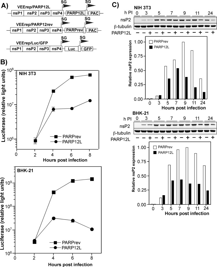
Alphaviruses represent a highly important group of human and animal pathogens, which are transmitted by mosquito vectors between vertebrate hosts. The hallmark of alphavirus infection in vertebrates is the induction of a high-titer viremia, which is strongly dependent on the ability of the virus to interfere with host antiviral responses on both cellular and organismal levels. The identification of cellular factors, which are critical in orchestrating virus clearance without the development of cytopathic effect, may prove crucial in the design of new and highly effective antiviral treatments. To address this issue, we have developed a noncytopathic Venezuelan equine encephalitis virus (VEEV) mutant that can persistently replicate in cells defective in type I interferon (IFN) production or signaling but is cleared from IFN signaling-competent cells. Using this mutant, we analyzed (i) the spectrum of cellular genes activated by virus replication in the persistently infected cells and (ii) the spectrum of genes activated during noncytopathic virus clearance. By applying microarray-based technology and bioinformatic analysis, we identified a number of IFN-stimulated genes (ISGs) specifically activated during VEEV clearance. One of these gene products, the long isoform of PARP12 (PARP12L), demonstrated an inhibitory effect on the replication of VEEV, as well as other alphaviruses and several different types of other RNA viruses. Additionally, overexpression of two other members of the PARP gene superfamily was also shown to be capable of inhibiting VEEV replication.


Figures

Fig 1
Fig 1
The VEEV variant with a mutated capsid gene persistently replicates in IFN-α/βR−/− MEFs but is cleared from IFN-competent cells. (A) Schematic representation of the VEEV genomes encoding wt and mutated capsid proteins. The mutated amino acids are presented in red. The NLS and the carboxy terminus of the supra-NES are indicated by boxes (3). Underlined amino acids indicate connecting peptide. nsP1 to -4 indicate nonstructural genes. C, E2, and E1 indicate structural genes. SG indicates the subgenomic RNA promoter. (B) NIH 3T3 cells and IFN-α/βR−/− MEFs were infected with the indicated viruses at an MOI of 20 PFU/cell. Media were replaced at the indicated time points, and titers of the released viruses were measured by plaque assay on BHK-21 cells. (C) Concentrations of IFN-β were measured in the samples (see panel B) harvested from the infected NIH 3T3 cells.
Fig 2
Fig 2
Microarray data analysis. Panels A, B, and C present the sequential analysis used to reduce the number of genes to analyze further in this study (see the text for details). In each, the first panel shows the gene expression profiles in the VEEV/GFP/C1-infected NIH 3T3 cells. The expression profiles of the same genes in IFN-β-treated NIH 3T3 cells and in the infected IFN-α/βR−/− MEFs are presented in the second and the third panels, respectively. Each line reflects expression levels of a single gene at different time points. The red lines represent the genes whose expression was activated the most, and the blue color indicates the genes whose expression was downregulated the most in the VEEV/GFP/C1-infected compared to mock-infected NIH 3T3 cells. Other colors indicate intermediate levels of activation and downregulation of gene expression. The arrow indicates the end of IFN-β treatment.
Fig 3
Fig 3
Four cellular genes were selected for further analysis. (A) Scheme used to define the final set of genes with putative antiviral functions. (B) Expression profiles of cellular genes that were selected for testing of their antiviral activities.
Fig 4
Fig 4
qPCR analysis confirms the microarray data. The selected genes demonstrate higher levels of activation in RT-qPCR tests than those detected in the microarray experiments. The experiments were performed in triplicates (see Materials and Methods for details).
Fig 5
Fig 5
PARP12L and Bst2 expression has a negative effect on VEEV mutant replication. (A) Schematic representation of recombinant viral genomes. HG indicates a position of cloned heterologous genes. (B) Schematic representation of long and short isoforms of PARP12 protein. (C) Replication of recombinant VEEV variants expressing the indicated heterologous genes in NIH 3T3 cells. Cells were infected at an MOI of 10 PFU/cell, media were replaced at the indicated times, and virus titers were determined by plaque assay on BHK-21 cells.
Fig 6
Fig 6
The antiviral effects of PARP12L and Bst2, expressed from the viral genome, is dependent on the applied MOI and cell type. (A) The schematic representation of recombinant VEEV genomes encoding mutated capsid protein, Bst2 and the PARP12L genes in direct or reverse orientations. (B) NIH 3T3 and BHK-21 cells were infected with recombinant viruses encoding PARP12L, Bst2, or PARP12L in reverse orientation at the indicated MOIs. Media were harvested at the indicated times postinfection, and virus titers were determined by plaque assay on BHK-21 cells.
Fig 7
Fig 7
Expression of PARP12L affects replication of VEEV encoding wt capsid protein. (A) Schematic representation of recombinant VEEV genomes encoding wt capsid protein and the PARP12L genes in direct or reverse orientations. (B) Analysis of replication of the recombinant viruses in BHK-21 and NIH 3T3 cells. Media were harvested at different times postinfection, and virus titers were determined by plaque assay on BHK-21 cells.
Fig 8
Fig 8
Expression of PARP12L affects replication of different alphaviruses and other RNA viruses. (A) Schematic representation of the VEEV replicon expressing the PARP12L gene, the control VEEV replicon, and the genomes of VEEV, SINV, and CHIKV used for superinfection. PAC indicates a puromycin acetyltransferase gene. (B and C) BHK-21 cells were infected with packaged VEEV replicons encoding PARP12L in either the direct or reverse orientation at an MOI of 20 infectious units/cell. One hour later, they were infected with the indicated viruses at an MOI of 1 PFU/cell, and media were replaced at the indicated times. Titers were determined by plaque assay on BHK-21 cells for all viruses.
Fig 9
Fig 9
Expression of PARP12L strongly affects expression of genes carried by the recombinant VEEV genome. (A) Schematic representation of VEEV replicons encoding PARP12L in either the direct or reverse orientation or firefly luciferase (Luc). (B) NIH 3T3 and BHK-21 cells were infected with packaged VEEV replicons expressing PARPrev and PARP12L at an MOI of 20 (inf.u./cell). After incubation for 1 h at 37°C, they were superinfected with packaged VEErep/Luc/GFP replicon at an MOI of 1 (inf.u./cell). Luciferase activity was measured at the indicated time points. (C) BHK-21 and NIH 3T3 cells were infected with packaged VEErep/PARP12L or VEErep/PARPrev replicons at an MOI of 20. Intracellular accumulation of VEEV nsP2 was measured at the indicated time points by Western blotting as described in Materials and Methods. The data were normalized to the level of β-tubulin.
Fig 10
Fig 10
Expression of some other PARP proteins also downregulates VEEV replication. (A) Schematic representations of the VEEV genome encoding different PARP proteins and of different PARPs used in this study. Zn fingers and domains are indicated. (B) Activation of different PARP genes in NIH 3T3 cells infected with VEEV/GFP/C1 or treated with IFN-β for 24 h. Activation of these genes was measured in the microarray experiments presented in Fig. 2 and 3. (C) Replication of recombinant PARP-expressing viruses and VEEV/PARPrev in BHK-21 and NIH 3T3 cells after infection at an MOI of 10 PFU/cell. Media were replaced at the indicated time points, and virus titers were determined by plaque assay on BHK-21 cells.

References

    1. Aguilar PV, Leung LW, Wang E, Weaver SC, Basler CF. 2008. A five-amino-acid deletion of the eastern equine encephalitis virus capsid protein attenuates replication in mammalian systems but not in mosquito cells. J. Virol. 82:6972–6983 - PMC - PubMed
    1. Aguilar PV, Weaver SC, Basler CF. 2007. Capsid protein of eastern equine encephalitis virus inhibits host cell gene expression. J. Virol. 81:3866–3876 - PMC - PubMed
    1. Atasheva S, Fish A, Fornerod M, Frolova EI. 2010. Venezuelan equine encephalitis virus capsid protein forms a tetrameric complex with CRM1 and importin alpha/beta that obstructs nuclear pore complex function. J. Virol. 84:4158–4171 - PMC - PubMed
    1. Atasheva S, Garmashova N, Frolov I, Frolova E. 2008. Venezuelan equine encephalitis virus capsid protein inhibits nuclear import in mammalian but not in mosquito cells. J. Virol. 82:4028–4041 - PMC - PubMed
    1. Atasheva S, Krendelchtchikova V, Liopo A, Frolova E, Frolov I. 2010. Interplay of acute and persistent infections caused by Venezuelan equine encephalitis virus encoding mutated capsid protein. J. Virol. 84:10004–10015 - PMC - PubMed

Publication types

Substances

LinkOut - more resources