. 2012 Aug;86(16):8388-401.
doi: 10.1128/JVI.00612-12. Epub 2012 May 23.

Suppression of the interferon and NF-κB responses by severe fever with thrombocytopenia syndrome virus

Affiliations

Suppression of the interferon and NF-κB responses by severe fever with thrombocytopenia syndrome virus

Bingqian Qu et al. J Virol. 2012 Aug.

Abstract

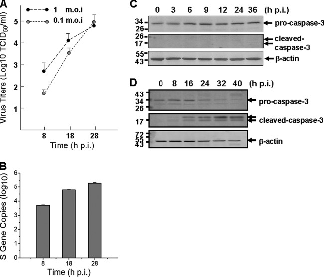
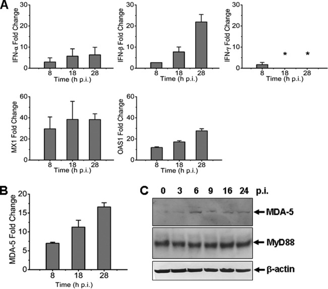
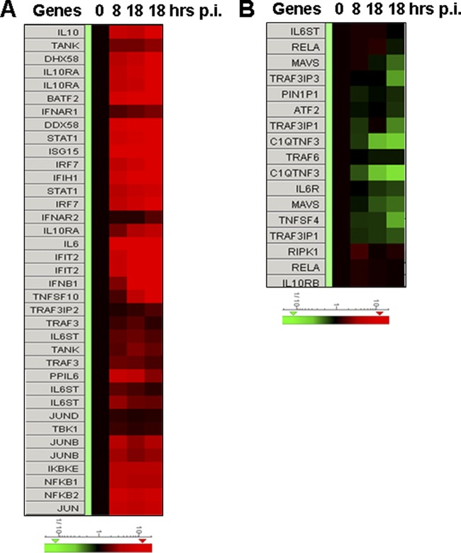
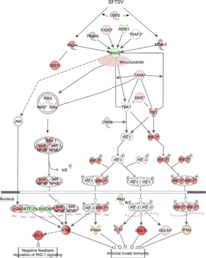
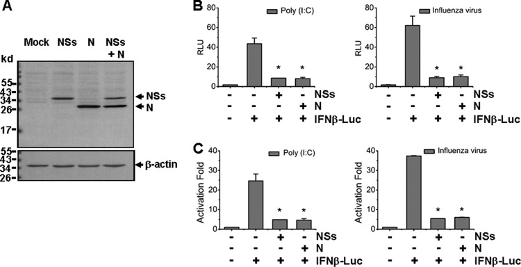
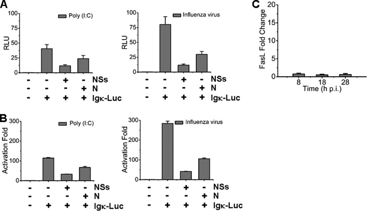
Severe fever with thrombocytopenia syndrome (SFTS) is an emerging infectious disease characterized by high fever, thrombocytopenia, multiorgan dysfunction, and a high fatality rate between 12 and 30%. It is caused by SFTS virus (SFTSV), a novel Phlebovirus in family Bunyaviridae. Although the viral pathogenesis remains largely unknown, hemopoietic cells appear to be targeted by the virus. In this study we report that human monocytes were susceptible to SFTSV, which replicated efficiently, as shown by an immunofluorescence assay and real-time reverse transcription-PCR. We examined host responses in the infected cells and found that antiviral interferon (IFN) and IFN-inducible proteins were induced upon infection. However, our data also indicated that downregulation of key molecules such as mitochondrial antiviral signaling protein (MAVS) or weakened activation of interferon regulatory factor (IRF) and NF-κB responses may contribute to a restricted innate immunity against the infection. NSs, the nonstructural protein encoded by the S segment, suppressed the beta interferon (IFN-β) and NF-κB promoter activities, although NF-κB activation appears to facilitate SFTSV replication in human monocytes. NSs was found to be associated with TBK1 and may inhibit the activation of downstream IRF and NF-κB signaling through this interaction. Interestingly, we demonstrated that the nucleoprotein (N), also encoded by the S segment, exhibited a suppressive effect on the activation of IFN-β and NF-κB signaling as well. Infected monocytes, mainly intact and free of apoptosis, may likely be implicated in persistent viral infection, spreading the virus to the circulation and causing primary viremia. Our findings provide the first evidence in dissecting the host responses in monocytes and understanding viral pathogenesis in humans infected with a novel deadly Bunyavirus.


Figures

Fig 1
Fig 1
Human THP-1 monocytes were susceptible to SFTSV infection. (A) THP-1 cells were mock infected or infected with SFTSV. More adhered cells were observed at 18 and 38 hpi in infected cells (magnification, ×100). Uninfected and infected cells were washed with PBS three times at different time points. (B) IFA for SFTSV antigens in infected THP-1 cells. The cells were mock infected or infected with the virus and fixed at 18 or 28 hpi. After permeabilization with Triton X-100, the cells were incubated with a human anti-SFTSV serum at a dilution of 1:100, followed by a staining with FITC anti-human IgG. (C) Percentage of THP-1 cells during the course of infection was positive with viral antigens at various time points postinfection. (D) IFA for SFTSV antigens in infected Vero cells.
Fig 2
Fig 2
Replication of SFTSV in human monocytes without causing apoptosis. (A) Replicative curve of SFTSV in the culture media of infected THP-1 cells. Culture media of infected THP-1 cells were taken at 8, 18, and 28 hpi, serially diluted, and inoculated in Vero cells. Infectious virus titers were determined after the cells were examined with IFA and calculated based on the Reed and Muench method. (B) Efficient replication of SFTSV in monocytes. Total RNA were extracted from either control or infected THP-1 cells at 8, 18, and 28 hpi and reverse transcribed before cDNAs were subjected to real-time PCR and the S gene copies were quantified. (C) No apoptosis was induced during the infection. Western blot analysis was performed to determine that no cleavage/activation of caspase-3 occurred in SFTSV-infected cells. (D) Apoptotic control. Caspase-3 was cleaved and activated in etoposide (10 μM)-treated THP-1 cells.
Fig 3
Fig 3
Antiviral responses induced in SFTSV-infected human monocytes. Total RNA were prepared from noninfected and infected cells at various time points postinfection, and real-time RT-PCR was performed to measure the transcript levels of IFN-α, IFN-β, IFN-γ, MX1, and OAS1 (A) and MDA-5 (B) at 8, 18, and 28 hpi, respectively. (C) Induction of MDA-5 expression. Cell lysates were prepared and subjected to SDS-PAGE and Western blot analysis with antibodies against MDA-5, MyD88, and β-actin at the time points as indicated.
Fig 4
Fig 4
Heat maps showing the expression profiles of genes in IFN and NF-κB signaling pathways in SFTSV-infected monocytes. Total RNA from noninfected and infected cells at 8, 18, and 28 hpi were prepared, and cRNA was labeled with Cy3-CTP before they were subjected to hybridization on A4x44 microarray slides (Agilent) and scanning. The GeneData Expressionist platform was used to calculate the relative fold changes of genes that are involved in IFN and NF-κB responses (A and B). Log ratios are depicted in red (upregulated) or green (downregulated).
Fig 5
Fig 5
NF-κB signaling analysis in THP-1 cells infected with SFTSV. The IPA platform was used to analyze components involved in the NF-κB response and their relationships and interactions. Fold changes of transcript expression levels of the genes in NF-κB signaling at 18 hpi were imported from the Agilent microarray analysis described above. Fold changes are shown in red (upregulation) and green (downregulation).
Fig 6
Fig 6
Pathway analysis of the antiviral IFN response in infected THP-1 cells. The IPA platform was used to analyze components of the IFN response involved in both IRF and NF-κB signaling. Fold changes of transcript expression levels of the genes in IRF and NF-κB signaling at 18 hpi were imported from the Agilent microarray analysis. Fold changes are shown in red (upregulation) and green (downregulation).
Fig 7
Fig 7
NSs and N proteins inhibited IFN-β promoter activity. (A) HEK293T cells were transfected with the plasmids expressing NSs, N, or both, and Western blot analysis was performed to examine the expression levels of NSs and N proteins. (B and C) IFN-β promoter activity was suppressed by NSs and N proteins in a luciferase assay. Cells were cotransfected with pRK5-NSs or pRK5-N plasmid, along with pGL3-IFNβ-Luc and pRL8 for 24 h, followed by stimulation with 50 μg of poly(I:C)/ml or infection with 0.5 MOI of an avian influenza virus for another 6 h. Cell lysates were subjected to lysis and the lysates were measured for luciferase activities. The results are shown as RLU (B) and activation fold (C).
Fig 8
Fig 8
NSs and N proteins suppressed NF-κB promoter activity. HEK293T cells were cotransfected with plasmids expressing NSs or N, along with pGL3-Igκ-Luc and pRL8, followed by stimulation with 50 μg of poly(I:C)/ml or 0.5 MOI of the avian influenza virus for additional 6 h. The results of luciferase activities were analyzed as RLU (A) and activation fold (B). (C) Transcript expression levels of FasL in infected cells were determined by real-time RT-PCR.
Fig 9
Fig 9
NSs association with TBK1 and transient NF-κB activation. (A) Coimmunoprecipitation of NSs and TBK1. Pretreated cell lysates in pRK5-Flag-NSs-transfected HEK293 were immunoprecipitated with the anti-Flag antibody. The immunoprecipitates were denatured and subjected to SDS-PAGE and Western blot analyses with anti-Flag and anti-TBK1 antibodies, respectively. (B) Transient phosphorylation and activation of NF-κB p65 (RelA) and NF-κB2, and degradation of IκB-α in infected THP-1 cells. Cell lysates at various time points postinfection were prepared from noninfected or infected cells, and subsequently subjected to SDS-PAGE and Western blot analyses. (C) Inhibition of NF-κB signaling suppressed SFTSV replication. THP-1 cells were pretreated with inhibitors of NF-κB (100 nM) or IKK (10 μM) for 1 h, and subsequently infected with SFTSV. At 24 hpi, total RNA was prepared for reverse transcription and cDNA were subjected to real-time PCR for quantifying S gene copies in the infected cells. (D) Infectious virus titers in NF-κB inhibitor-treated THP-1 cells were lower than those in nontreated cells. Culture media from infected THP-1 cells, treated or not treated with the inhibitor, were diluted and subsequently used to infect Vero cells in 12-well plates for infectious virus titration (TCID50, Student t test; *, P < 0.05).

Similar articles

Cited by

References

    1. Bachmann M, Moroy T. 2005. The serine/threonine kinase Pim-1. Int. J. Biochem. Cell Biol. 37:726–730 - PubMed
    1. Bahram S, Arnold D, Bresnahan M, Strominger JL, Spies T. 1991. Two putative subunits of a peptide pump encoded in the human major histocompatibility complex class II region. Proc. Natl. Acad. Sci. U. S. A. 88:10094–10098 - PMC - PubMed
    1. Bao CJ, et al. 2007. A family cluster of infections by a newly recognized bunyavirus in eastern China: further evidence of person-to-person transmission. Clin. Infect. Dis. 53:1208–1214 - PubMed
    1. Billecocq A, et al. 2004. NSs protein of Rift Valley fever virus blocks interferon production by inhibiting host gene transcription. J. Virol. 78:9798–9806 - PMC - PubMed
    1. Bishop DH. 1986. Ambisense RNA genomes of arenaviruses and phleboviruses. Adv. Virus Res. 31:1–51 - PMC - PubMed

Publication types

LinkOut - more resources