

Targeting bladder tumor cells in voided urine of Chinese patients with FITC-CSNRDARRC peptide ligand
- PMID: 22623877
- PMCID: PMC3358809
- DOI: 10.2147/OTT.S31368
Targeting bladder tumor cells in voided urine of Chinese patients with FITC-CSNRDARRC peptide ligand
Abstract
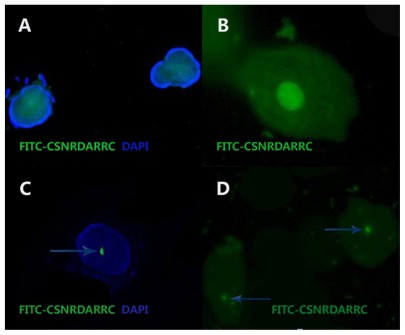
Objective: To study the practicality of the FITC-CSNRDARRC peptide ligand (containing the Cys-Ser-Asn-Arg-Asp-Ala-Arg-Arg-Cys nonapeptide) in diagnosing and monitoring bladder tumors.
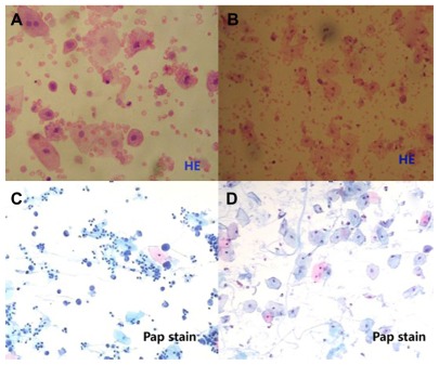
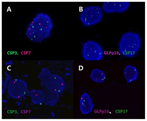
Materials and methods: Between March 2011 and September 2011, 80 consecutive patients with radiographic abnormalities, localizing hematuria, other symptoms, or signs were studied using the FITC-CSNRDARRC ligand, urinary cytology (UC), and fluorescence in situ hybridization (FISH). The sensitivity and specificity of these three technologies were determined and compared. Cystoscopy and tissue biopsy were taken as the "gold standards" for bladder tumor diagnosis in this study.
Results: Twenty-nine out of 80 patients were diagnosed with a bladder tumor via histopathological examination. The FITC-CSNRDARRC ligand was positive in 23 out of 29 bladder tumor patients and produced false negatives in six (20.69%) patients. The UC was positive in six out of 29 bladder tumor patients and produced false negatives in 23 (79.31%) patients. The FISH was positive in 21 out of 29 bladder tumor patients and produced false negatives in eight (27.59%) patients. The overall sensitivity as verified by the FITC-CSNRDARRC ligand was much higher than in UC (79.31% versus 20.69%, P < 0.001) and was slightly higher than in FISH (79.31% versus 72.41%, P = 0.625). The sensitivity of FISH was significantly higher than that of UC (72.41% versus 20.69%, P < 0.001). Sensitivities of the FITC-CSNRDARRC ligand and UC by grade were 58.33% versus 8.3% for low-grade (LG) tumors (P = 0.031) and 94.12% versus 29.41% for high-grade (HG) tumors (P = 0.003), respectively. The advantage was maintained in terms of the detection of invasive tumors between the FITC-CSNRDARRC ligand and UC (90.48% versus 23.81%, P = 0.001) as well as between FISH and UC (85.71% versus 23.81%, P = 0.003). The specificities for the FITC-CSNRDARRC ligand, UC, and FISH were 100%.
Conclusion: Results show that the FITC-CSNRDARRC ligand is a promising noninvasive tool for diagnosis and surveillance in patients suspected of having a new bladder tumor.
Keywords: FITC-CSNRDARRC ligand; bladder tumor; fluorescent probe; tumor-targeting.
Figures
Similar articles
-
[Numerical aberrations of chromosomes 11 and 17 detected by fish--fluorescence in situ hybridization combined with cytology in exfoliated cells from voided urine in patients with urothelial carcinoma of the bladder].Harefuah. 2007 Dec;146(12):914-9, 1000. Harefuah. 2007. PMID: 18254439 Hebrew.
-
Prospective evaluation of fluorescence in situ hybridization for diagnosing urothelial carcinoma.Oncol Lett. 2017 May;13(5):3928-3934. doi: 10.3892/ol.2017.5926. Epub 2017 Mar 27. Oncol Lett. 2017. PMID: 28529600 Free PMC article.
-
Predictive value of MCM5 (ADXBLADDER) analysis in urine of men evaluated for the initial diagnosis of bladder cancer: A comparative prospective study.Diagn Cytopathol. 2020 Nov;48(11):1034-1040. doi: 10.1002/dc.24530. Epub 2020 Jun 20. Diagn Cytopathol. 2020. PMID: 32562513
-
DNA-based molecular cytology for bladder cancer surveillance.Urology. 2006 Mar;67(3 Suppl 1):35-45; discussion 45-7. doi: 10.1016/j.urology.2006.01.039. Urology. 2006. PMID: 16530074 Review.
-
Detection of bladder tumors by immunostaining of the Lewis X antigen in cells from voided urine.Urology. 1995 Aug;46(2):173-7. doi: 10.1016/s0090-4295(99)80189-7. Urology. 1995. PMID: 7624989 Review.
Cited by
-
Multicomponent, peptide-targeted glycol chitosan nanoparticles containing ferrimagnetic iron oxide nanocubes for bladder cancer multimodal imaging.Int J Nanomedicine. 2016 Aug 29;11:4141-55. doi: 10.2147/IJN.S109494. eCollection 2016. Int J Nanomedicine. 2016. PMID: 27621615 Free PMC article.
-
Combinatorial peptide libraries: mining for cell-binding peptides.Chem Rev. 2014 Jan 22;114(2):1020-81. doi: 10.1021/cr400166n. Epub 2013 Dec 3. Chem Rev. 2014. PMID: 24299061 Free PMC article. Review. No abstract available.
-
Polydopamine and peptide decorated doxorubicin-loaded mesoporous silica nanoparticles as a targeted drug delivery system for bladder cancer therapy.Drug Deliv. 2017 Nov;24(1):681-691. doi: 10.1080/10717544.2017.1309475. Drug Deliv. 2017. PMID: 28414557 Free PMC article.
-
Application of Bld-1-Embedded Elastin-Like Polypeptides in Tumor Targeting.Sci Rep. 2018 Mar 1;8(1):3892. doi: 10.1038/s41598-018-21910-z. Sci Rep. 2018. PMID: 29497090 Free PMC article.
References
-
- Yoo JH, Suh B, Park TS, et al. Analysis of fluorescence in situ hybridization, mtDNA quantification, and mtDNA sequence for the detection of early bladder cancer. Cancer Genet and Cytogenet. 2010;198(2):107–117. - PubMed
-
- Tomera KM. NMP22 (R) BladderChek (R) Test: Point-of-care technology with life and money-saving potential. Expert Rev Mol Diagn. 2004;4(6):783–794. - PubMed
-
- Sylvester RJ, van der Meijden APM, Oosterlinck W, et al. Predicting recurrence and progression in individual patients with stage Ta T1 bladder cancer using EORTC risk tables: a combined analysis of 2596 patients from seven EORTC trials. Eur Urol. 2006;49(3):466–477. - PubMed
LinkOut - more resources
Full Text Sources
