. 2012 May 16:6:112.
doi: 10.3389/fnhum.2012.00112. eCollection 2012.

Correlated components of ongoing EEG point to emotionally laden attention - a possible marker of engagement?

Affiliations

Correlated components of ongoing EEG point to emotionally laden attention - a possible marker of engagement?

Jacek P Dmochowski et al. Front Hum Neurosci. .

Abstract

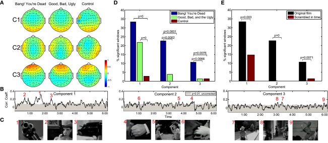
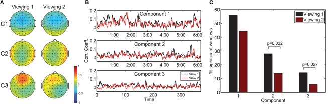
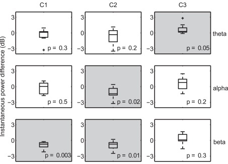
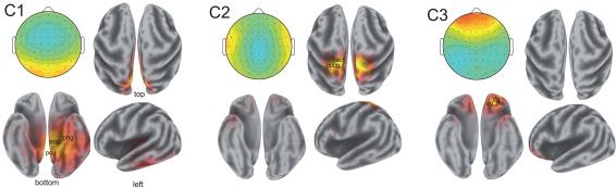
Recent evidence from functional magnetic resonance imaging suggests that cortical hemodynamic responses coincide in different subjects experiencing a common naturalistic stimulus. Here we utilize neural responses in the electroencephalogram (EEG) evoked by multiple presentations of short film clips to index brain states marked by high levels of correlation within and across subjects. We formulate a novel signal decomposition method which extracts maximally correlated signal components from multiple EEG records. The resulting components capture correlations down to a one-second time resolution, thus revealing that peak correlations of neural activity across viewings can occur in remarkable correspondence with arousing moments of the film. Moreover, a significant reduction in neural correlation occurs upon a second viewing of the film or when the narrative is disrupted by presenting its scenes scrambled in time. We also probe oscillatory brain activity during periods of heightened correlation, and observe during such times a significant increase in the theta band for a frontal component and reductions in the alpha and beta frequency bands for parietal and occipital components. Low-resolution EEG tomography of these components suggests that the correlated neural activity is consistent with sources in the cingulate and orbitofrontal cortices. Put together, these results suggest that the observed synchrony reflects attention- and emotion-modulated cortical processing which may be decoded with high temporal resolution by extracting maximally correlated components of neural activity.

Keywords: attention; brain reading; canonical correlation analysis; electroencephalography; engagement.


Figures

Figure 1
Figure 1
Neural correlations during two critically acclaimed films and one amateur control. The scalp projections of the first three maximally correlated components show a remarkable congruence across the three films shown (A). The within-subject correlation peaks at particularly arousing moments of “Bang! You’re Dead,” (B,C) with over 30% of the film resulting in statistically significant correlations in the first component (D). On the other hand, any extended periods of statistically significant correlation fail to arise during the control clip. Moreover, when presenting “Bang! You’re Dead” with its scenes scrambled in time, a significant reduction in neural correlation ensues (E).
Figure 2
Figure 2
The effect of prior exposure on neural correlation. The scalp projections of the components maximizing population ISC during the first viewing are largely congruent to those stemming from viewing 2 (A). However, the resulting time-resolved correlation measures are significantly lower during the second viewing (B). Furthermore, more time windows exhibit statistically significant ISC in the first viewing (C).
Figure 3
Figure 3
Differences in instantaneous power during moments of high versus low neural correlation. Distributions are constructed along the subject dimension (n = 10, with statistically significant effects denoted with a light gray background). High correlation windows are marked by synchronization of theta activity in the third component, desynchronization of alpha in the second component, and desynchronization of beta in the first and second components.
Figure 4
Figure 4
Sources of correlated neural activity for components 1, 2, and 3. The scalp projections of the correlated activity are shown in the top left of each pane; the estimated distributions of cortical sources are depicted in the remaining three panes. The correlated activity of component 1 suggests involvement of the posterior cingulate gyrus (Brodmann Area 31, labeled pcg), the parahippocampal gyrus (Brodmann Area 27, phg), and precuneus (Brodmann Area 7, pcu). The postcentral gyrus (pocg) and paracentral lobule (pacl) are implicated in the localization of the activity in component two. The activity captured by component 3 is consistent with sources in the inferior frontal gyrus (ifg) and the orbital gyrus (og). Anatomical locations shown are approximate.

References

    1. Anderson A. K., Wais P. E., Gabrieli J. D. E. (2006). Emotion enhances remembrance of neutral events past. Proc. Natl. Acad. Sci. U.S.A. 103, 1599–160410.1073/pnas.0509537103 - DOI - PMC - PubMed
    1. Ariely D., Berns G. S. (2010). Neuromarketing: the hope and hype of neuroimaging in business. Nat. Rev. Neurosci. 11, 284–29210.1038/nrn2795 - DOI - PMC - PubMed
    1. Bellman R. (1957). Dynamic Programming. Princeton: Princeton University Press
    1. Benjamini Y., Hochberg Y. (1995). Controlling the false discovery rate: a practical and powerful approach to multiple testing. J. R. Stat. Soc. Series B Stat. Methodol. 57, 289–300
    1. Boashash B. (1992). Estimating and interpreting the instantaneous frequency of a signal – part 1: fundamentals. Proc. IEEE 80, 520–53810.1109/5.135376 - DOI

LinkOut - more resources