. 2012;7(5):e37004.
doi: 10.1371/journal.pone.0037004. Epub 2012 May 18.

A scalable system for production of functional pancreatic progenitors from human embryonic stem cells

Affiliations

A scalable system for production of functional pancreatic progenitors from human embryonic stem cells

Thomas C Schulz et al. PLoS One. 2012.

Abstract

Development of a human embryonic stem cell (hESC)-based therapy for type 1 diabetes will require the translation of proof-of-principle concepts into a scalable, controlled, and regulated cell manufacturing process. We have previously demonstrated that hESC can be directed to differentiate into pancreatic progenitors that mature into functional glucose-responsive, insulin-secreting cells in vivo. In this study we describe hESC expansion and banking methods and a suspension-based differentiation system, which together underpin an integrated scalable manufacturing process for producing pancreatic progenitors. This system has been optimized for the CyT49 cell line. Accordingly, qualified large-scale single-cell master and working cGMP cell banks of CyT49 have been generated to provide a virtually unlimited starting resource for manufacturing. Upon thaw from these banks, we expanded CyT49 for two weeks in an adherent culture format that achieves 50-100 fold expansion per week. Undifferentiated CyT49 were then aggregated into clusters in dynamic rotational suspension culture, followed by differentiation en masse for two weeks with a four-stage protocol. Numerous scaled differentiation runs generated reproducible and defined population compositions highly enriched for pancreatic cell lineages, as shown by examining mRNA expression at each stage of differentiation and flow cytometry of the final population. Islet-like tissue containing glucose-responsive, insulin-secreting cells was generated upon implantation into mice. By four- to five-months post-engraftment, mature neo-pancreatic tissue was sufficient to protect against streptozotocin (STZ)-induced hyperglycemia. In summary, we have developed a tractable manufacturing process for the generation of functional pancreatic progenitors from hESC on a scale amenable to clinical entry.


Conflict of interest statement

Competing Interests: The indicated authors are employees or former employees of Viacyte, receiving salary and stock options. The paper describes or references intellectual property owned by Viacyte ([1] Green C, Yu X, Bang A, Brandon E, Kelly O, Agulnick A, Baetge E, D’Amour K, Schulz TC, Robins A. Stem cell aggregate suspension compositions and methods of differentiation thereof. US Patent 8,008,075. [2] Robins A, Schulz, T. Methods and compositions for feeder-free pluripotent stem cell media containing human serum. US Patent application 20100311164. [3] Robins A, Schulz T. Compositions and methods useful for culturing differentiable cells. US Patent application 20080268534), and a third-party product that generates royalty income (LifeTechnologies: StemPro hESC SFM). This does not alter the authors' adherence to all the PLoS ONE policies on sharing data and materials.

Figures

Figure 1
Figure 1. Directed pancreatic differentiation of CyT49 in suspension culture.
(A) Schematic representation of aggregation and the four-stage differentiation protocol from hESC (ES) to mesendoderm (ME), definitive endoderm (DE), primitive gut tube (PG), posterior foregut (PF), and a mixed population comprising primarily of pancreatic endoderm (PE) and endocrine precursor/endocrine cells (EP). Culture conditions, timing and rotation speeds are indicated. Markers used to identify the different stages are shown.*TBI: TGF-β RI kinase Inhibitor IV, first day of Stage-2 only. **ITS: Insulin-Transferrin-Selenium used at different concentrations in Stage-1 and -2. HrgB, heregulin 1β; D/F12, DMEM/F12; TT, TTNPB; CYC, cyclopamine; NOG, noggin. (B,C,D) Hematoxylin and eosin staining of sections of CyT49 aggregates after (B) culture in StemPro medium for 2 days (paraffin section), or (C) at d5, or (D) at d12 of differentiation (frozen sections). Scale bars, 50 µm. (E) Immunofluorescence analysis of sections of d5 aggregates stained with OCT4/DAPI, or (F) FOXA2/DAPI. A single cluster of ∼four OCT4+ nuclei within the field of view is indicated (arrow). Imaged with a 20× objective. Immunofluorescence analysis of d12 aggregates for (G) NKX6-1, PDX1 and CHGA expression, and (H) FOXA2, PDX1 and NKX2-2 expression. Imaged with a 40× objective. DAPI, 4′,6-diamidino-2-phenylindole.
Figure 2
Figure 2. Cellular composition of pancreatic differentiation runs.
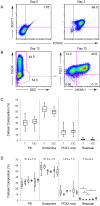
(A) Flow cytometric analyses of representative d0 undifferentiated (left), and d2 DE populations (right), co-stained with anti-SOX17 and anti-FOXA2. The percentages of total intact double-positive cells are indicated. (B) Flow cytometric analysis of a d12 differentiated population (scaled differentiation run, Expt #21) co-stained with anti-CHGA, anti-NKX6-1 and anti-PDX1. The analyses were performed by first gating on the CHGA- population, then plotting NKX6-1/PDX1 expression. Percentages of total intact cells for each cell subset are indicated. (C) The cellular composition of PE (CHGA−/NKX6-1+/PDX1+/−), endocrine (CHGA+/NKX6-1+/−/PDX1+/−), PDX1-only endoderm (CHGA−/NKX6-1−/PDX1+) and residual (CHGA−/NKX6-1−/PDX1) populations for all 37 scaled differentiation runs (T: total, n = 49 analyses) and the selected processes (13C: Table S2 Expt #18–21, 25–30, 35–37. n = 17 analyses) are plotted. The 13C group of differentiation runs were all performed within an 11 month period. The box plots show the median, second and third quartile (box), max and min values for each data set. The means within each group were not statistically different. (D) Pancreatic composition of differentiation runs arranged by CyT49 cell bank: (left to right) RCB-D (n = 19), MCB3 (n = 6), MCB4 (n = 7), MCB5 (n = 3), RCB-Dw (n = 11) and WCB4B (n = 3). The mean ± SEM (%) for each population is indicated at the top. Statistically significant differences are indicated (*: p<0.01).
Figure 3
Figure 3. Digital mRNA profiling of scaled pancreatic differentiation runs.
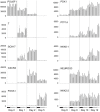
The dynamics of gene expression demonstrated that undifferentiated cells (POU5F1) were directed through mesendoderm (Brachyury: T), definitive endoderm (SOX17, CXCR4), primitive gut tube (FOXA1), posterior foregut (PDX1), to form pancreatic epithelium (NKX6-1, PTF1A), and endocrine cells (NEUROG3, NKX2-2). Precise temporal control and consistency between manufacturing runs indicated a reproducible and robust specification of each lineage. The plots are ordered according to CyT49 cell bank (left to right): black bar (MCB4: Expt #18–21), grey bar (RCB-Dw: Expt #25–30), open grey bar (WCB4B: Expt #35–37). The average and standard deviation of three biological replicates are plotted. Additional data is shown in Figs. S6, S7.
Figure 4
Figure 4. In vivo function of engrafted pancreatic differentiation runs.
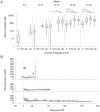
(A) GSIS response in high functioning engrafted animals (n = 166) at selected time points after implantation (weeks post-engraftment shown at top). Each time point shows F, fasting; 5 or 10 min; 30 min; and 60 min serum human C-peptide measurements. A significant increase in both fasting (p<0.01) and maximum GSIS (p<0.01) was observed from weeks 5–10 to weeks 11–50. Other statistically significant differences are indicated (*: p<0.01). The box plots show the median, second and third quartile (box), max and min values for serum human C-peptide (pM). A log scale is used to display both early and late time points. Empty plots indicate that no data were collected. Additional analyses and the number of ELISA samples for each data set are shown in Fig. S8 and Table S3, respectively. (B) Protection against STZ-mediated hyperglycemia in long-term grafts. Non-fasting blood glucose measurements (mg/dL) are plotted for three cohorts of mice (n = 15, one animal not shown) over days post-STZ treatment. STZ was administered on days 1–5 (black bar), in mice containing high-functioning grafts, 4–5 months after engraftment. Explanting grafts (arrow) led to hyperglycemia. The cohorts were derived from expt #34 (upper), #30 (middle) and #2 (bottom) (Table S2, banks RCB-D and RCB-Dw).
Figure 5
Figure 5. Histological and immunofluorescence analyses of CyT49-derived neo-pancreatic grafts at 18 weeks post-implant.
(A) Hematoxylin and eosin staining of a graft cross-section, and (B) higher magnification of boxed area. (C, D) GCG, SST and INS staining in a cross-section of a graft, demonstrating single-pancreatic hormone expression and large clusters of INS+ cells. (E) Co-expression of NKX6-1, PDX1 and INS. (F) TRY, INS and DAPI staining. (G) CK19, PDX1 and HuNU staining. GCG, glucagon; SST, somatostatin; INS, insulin; TRY, trypsin; HuNU, human nuclear antigen; CK19, cytokeratin 19. Grafts were from expt #18 (A, B), and #20 (C–G). These representative mice exhibited fasting human C-peptide levels of 229–865 pM, and maximum GSIS (30 or 60 min stimulation) of 2614–3485 pM at week 15. An additional ten representative grafts are shown in Figs. S10, S11, S12, S13, S14. Scale bars: 3 mm (A), 300 µm (B), 500 µm (C), 200 µm (D, F, G), 50 µm (E).
Figure 6
Figure 6. Schematic representation of the manufacturing process for pancreatic progenitors.
Scaled and high-density single-cell master (MCB) and working cell banks (WCB) of CyT49 cells were prepared with cGMP and serve as a virtually unlimited source of starting material for differentiation. Cryopreserved vials of 107 CyT49 cells from a qualified working cell bank are thawed and expanded in adherent culture conditions for 4 or 5 passages over a 2-week period. A single cell suspension is harvested and aggregated in rotational culture in 6-well trays. After 24 hrs the hESC aggregates are differentiated en masse with the 4-stage protocol to a population of pancreatic progenitors. For clinical development, scaled lots of differentiated aggregates will be produced with cGMP and cryopreserved, enabling a proportion of each lot to be tested for safety, efficacy and other regulatory considerations. Qualified lots of differentiated pancreatic progenitors will be thawed, recovered, formulated, loaded into a durable immunoisolation device and delivered for preclinical or clinical studies. The manufacturing process is amenable to further scaling via additional passages in adherent culture, as well as aggregation and differentiation in larger vessels.

References

    1. Shapiro AM, Lakey JR, Ryan EA, Korbutt GS, Toth E, et al. Islet transplantation in seven patients with type 1 diabetes mellitus using a glucocorticoid-free immunosuppressive regimen. N Engl J Med. 2000;343:230–238. - PubMed
    1. Matsumoto S. Clinical allogeneic and autologous islet cell transplantation: update. Diabetes Metab J. 2011;35:199–206. - PMC - PubMed
    1. Dominguez-Bendala J, Inverardi L, Ricordi C. Stem cell-derived islet cells for transplantation. Curr Opin Organ Transplant. 2011;16(1):76–82. - PMC - PubMed
    1. McCall MD, Toso C, Baetge EE, Shapiro AM. Are stem cells a cure for diabetes? Clin Sci (Lond) 2009;118:87–97. - PubMed
    1. D’Amour KA, Bang AG, Eliazer S, Kelly OG, Agulnick AD, et al. Production of pancreatic hormone-expressing endocrine cells from human embryonic stem cells. Nat Biotechnol. 2006;24:1392–1401. - PubMed

Publication types