Gut pathology and responses to the microsporidium Nosema ceranae in the honey bee Apis mellifera
- PMID: 22623972
- PMCID: PMC3356400
- DOI: 10.1371/journal.pone.0037017
Gut pathology and responses to the microsporidium Nosema ceranae in the honey bee Apis mellifera
Abstract
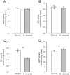
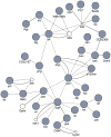
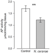
The microsporidium Nosema ceranae is a newly prevalent parasite of the European honey bee (Apis mellifera). Although this parasite is presently spreading across the world into its novel host, the mechanisms by it which affects the bees and how bees respond are not well understood. We therefore performed an extensive characterization of the parasite effects at the molecular level by using genetic and biochemical tools. The transcriptome modifications at the midgut level were characterized seven days post-infection with tiling microarrays. Then we tested the bee midgut response to infection by measuring activity of antioxidant and detoxification enzymes (superoxide dismutases, glutathione peroxidases, glutathione reductase, and glutathione-S-transferase). At the gene-expression level, the bee midgut responded to N. ceranae infection by an increase in oxidative stress concurrent with the generation of antioxidant enzymes, defense and protective response specifically observed in the gut of mammals and insects. However, at the enzymatic level, the protective response was not confirmed, with only glutathione-S-transferase exhibiting a higher activity in infected bees. The oxidative stress was associated with a higher transcription of sugar transporter in the gut. Finally, a dramatic effect of the microsporidia infection was the inhibition of genes involved in the homeostasis and renewal of intestinal tissues (Wnt signaling pathway), a phenomenon that was confirmed at the histological level. This tissue degeneration and prevention of gut epithelium renewal may explain early bee death. In conclusion, our integrated approach not only gives new insights into the pathological effects of N. ceranae and the bee gut response, but also demonstrate that the honey bee gut is an interesting model system for studying host defense responses.
Conflict of interest statement
Figures


References
-
- Becnel JJ, Andreadis TG. Microsporidia in insect. In: Wittner M, Weiss LM, editors. The Microsporidia and Microsporidiosis. Washington, DC: ASM Press; 1999. pp. 447–501.
-
- Higes M, Garcia-Palencia P, Martin-Hernandez R, Meana A. Experimental infection of Apis mellifera honeybees with Nosema ceranae (Microsporidia). J Invertebr Pathol. 2007;94:211–217. - PubMed
-
- Zander E. Tierische Parasiten als Krankenheitserreger bei der Biene. Münchener Bienenzeitung. 1909;31:196–204.
-
- Fries I, Feng F, Da Silva A, Slemeda SB, Pieniazek NJ. Nosema ceranae n. sp. (Microspora, Nosematidae), morphological and molecular characterization of a microsporidian parasite of the Asian honey bee Apis cerana (Hymenoptera, Apidae). Eur J Protistol. 1996;32:356–365.
-
- Fries I. Nosema ceranae in European honey bees (Apis mellifera). J Invertebr Pathol. 2010;103:S73–S79. - PubMed
Publication types
MeSH terms
Substances
LinkOut - more resources
Full Text Sources
Molecular Biology Databases

